Explore
 Validate
 Learn
Learn Western blot
 ELISA
ELISA Immunocytochemistry
Antibody data
- Antibody Data
- Antigen structure
- References [0]
- Comments [0]
- Validations
- Immunocytochemistry [1]
- Flow cytometry [1]
Submit
Validation data
Reference
Comment
Report error
- Product number
- MA5-42758 - Provider product page

- Provider
- Invitrogen Antibodies
- Product name
- CD70 Recombinant Rabbit Monoclonal Antibody (7F4S5)
- Antibody type
- Monoclonal
- Antigen
- Recombinant full-length protein
- Description
- Positive test controls include: Raji, 293T. Immunogen sequence: QRFAQAQQQL PLESLGWDVA ELQLNHTGPQ QDPRLYWQGG PALGRSFLHG PELDKGQLRI HRDGIYMVHI QVTLAICSST TASRHHPTTL AVGICSPASR SISLLRLSFH QGCTIASQRL TPLARGDTLC TNLTGTLLPS RNTDETFFGV QWVRP
- Reactivity
- Human
- Host
- Rabbit
- Isotype
- IgG
- Antibody clone number
- 7F4S5
- Vial size
- 100 μL
- Concentration
- 1 mg/mL
- Storage
- Store at 4°C short term. For long term storage, store at -20°C, avoiding freeze/thaw cycles.
No comments: Submit comment
Supportive validation
- Submitted by
- Invitrogen Antibodies (provider)
- Main image
- Experimental details
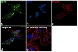
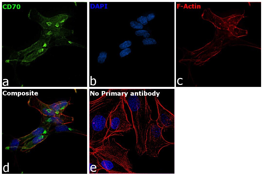
- Immunofluorescence analysis of CD70 was performed using 70% confluent log phase SK-O-V3 cells. The cells were fixed with 4% paraformaldehyde for 10 minutes, permeabilized with 0.1% Triton™ X-100 for 15 minutes, and blocked with 2% BSA for 45 minutes at room temperature. The cells were labeled with CD70 Recombinant Rabbit Monoclonal Antibody (7F4S5) (Product # MA5-42758, 1:100) in 0.1% BSA, incubated at 4 degree celsius overnight and then labeled with Goat anti-Rabbit IgG (Heavy chain), Superclonal™ Recombinant Secondary Antibody, Alexa Fluor™ 488 (Product # A27034, 1:2000) for 45 minutes at room temperature (Panel a: Green). Nuclei (Panel b:Blue) were stained with ProLong™ Diamond Antifade Mountant with DAPI (Product # P36962). F-actin (Panel c: Red) was stained with Rhodamine Phalloidin (Product # R415, 1:300). Panel d represents the merged image showing membrane and secretory localization. Panel e represents control cells with no primary antibody to assess background. The images were captured at 60X magnification.
Supportive validation
- Submitted by
- Invitrogen Antibodies (provider)
- Main image
- Experimental details
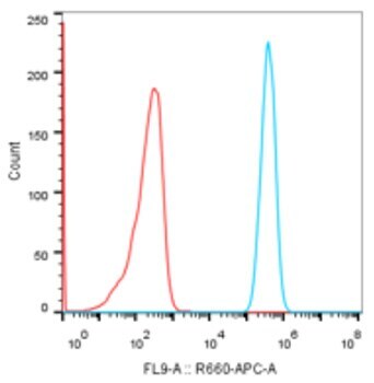
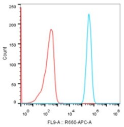
- Flow cytometry analysis of CD70 in Raji cells. Cells were stained with Rabbit IgG isotype control (1 μg/mL, Red line) or CD70 monoclonal antibody (Product # MA5-42758) (1 μg/mL, Blue line), followed by goat anti-Rabbit polyclonal antibody APC (1:200 dilution) staining.