Explore
 Validate
 Learn
Learn Western blot
Antibody data
- Antibody Data
- Antigen structure
- References [3]
- Comments [0]
- Validations
- Western blot [6]
- Immunocytochemistry [1]
- Immunoprecipitation [1]
- Immunohistochemistry [1]
Submit
Validation data
Reference
Comment
Report error
- Product number
- GTX104015 - Provider product page

- Provider
- GeneTex
- Proper citation
- GeneTex Cat#GTX104015, RRID:AB_1951155
- Product name
- Pyruvate Dehydrogenase E1 alpha antibody
- Antibody type
- Polyclonal
- Reactivity
- Human, Mouse, Rat
- Host
- Rabbit
Submitted references Critical Role of the Neonatal Fc Receptor (FcRn) in the Pathogenic Action of Antimitochondrial Autoantibodies Synergizing with Anti-desmoglein Autoantibodies in Pemphigus Vulgaris.
Protein kinase B (PKB/AKT1) formed signaling complexes with mitochondrial proteins and prevented glycolytic energy dysfunction in cultured cardiomyocytes during ischemia-reperfusion injury.
Skeletal muscle Nur77 expression enhances oxidative metabolism and substrate utilization.
Chen Y, Chernyavsky A, Webber RJ, Grando SA, Wang PH
The Journal of biological chemistry 2015 Sep 25;290(39):23826-37
The Journal of biological chemistry 2015 Sep 25;290(39):23826-37
Protein kinase B (PKB/AKT1) formed signaling complexes with mitochondrial proteins and prevented glycolytic energy dysfunction in cultured cardiomyocytes during ischemia-reperfusion injury.
Deng W, Leu HB, Chen Y, Chen YH, Epperson CM, Juang C, Wang PH
Endocrinology 2014 May;155(5):1618-28
Endocrinology 2014 May;155(5):1618-28
Skeletal muscle Nur77 expression enhances oxidative metabolism and substrate utilization.
Chao LC, Wroblewski K, Ilkayeva OR, Stevens RD, Bain J, Meyer GA, Schenk S, Martinez L, Vergnes L, Narkar VA, Drew BG, Hong C, Boyadjian R, Hevener AL, Evans RM, Reue K, Spencer MJ, Newgard CB, Tontonoz P
Journal of lipid research 2012 Dec;53(12):2610-9
Journal of lipid research 2012 Dec;53(12):2610-9
No comments: Submit comment
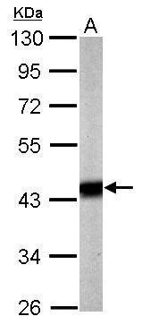
Supportive validation
- Submitted by
- GeneTex (provider)
- Main image

- Experimental details
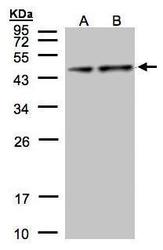
- Sample (20 ?g of whole cell lysate) A: mouse brain 10% SDS PAGE GTX104015 diluted at 1:5000 The HRP-conjugated anti-rabbit IgG antibody (GTX213110-01) was used to detect the primary antibody.
- Submitted by
- GeneTex (provider)
- Main image
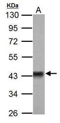
- Experimental details
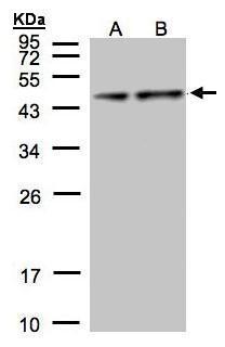
- Sample (50 ?g of whole cell lysate) A: Rat brain 10% SDS PAGE GTX104015 diluted at 1:3000 The HRP-conjugated anti-rabbit IgG antibody (GTX213110-01) was used to detect the primary antibody.
- Submitted by
- GeneTex (provider)
- Main image
- Experimental details
- Sample(30 ?g of whole cell lysate)A:Hep G2(GTX27900)B:MOLT4(GTX27912)12% SDS PAGEGTX104015 diluted at 1:1000The HRP-conjugated anti-rabbit IgG antibody (GTX213110-01) was used to detect the primary antibody.
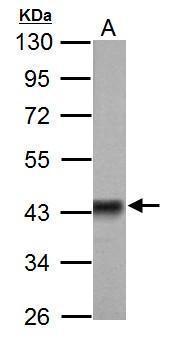
- Submitted by
- GeneTex (provider)
- Main image
- Experimental details
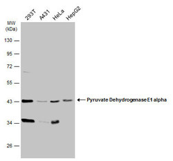
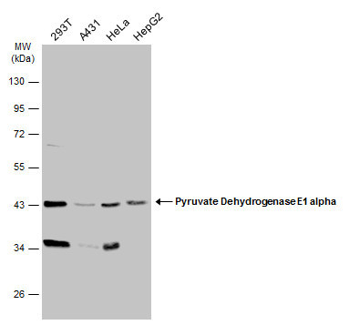
- Various whole cell extracts (30 ?g) were separated by 10% SDS-PAGE, and the membrane was blotted with Pyruvate Dehydrogenase E1 alpha antibody (GTX104015) diluted at 1:1000. The HRP-conjugated anti-rabbit IgG antibody (GTX213110-01) was used to detect the primary antibody.
- Submitted by
- GeneTex (provider)
- Main image
- Experimental details
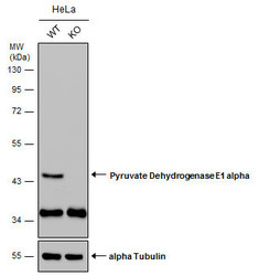
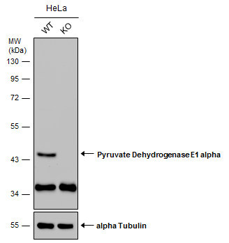
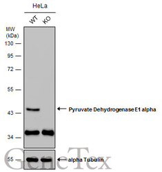
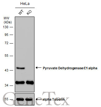
- Wild-type (WT) and Pyruvate Dehydrogenase E1 alpha knockout (KO) HeLa cell extracts (30 ?g) were separated by 10% SDS-PAGE, and the membrane was blotted with Pyruvate Dehydrogenase E1 alpha antibody (GTX104015) diluted at 1:1000. The HRP-conjugated anti-rabbit IgG antibody (GTX213110-01) was used to detect the primary antibody.
- Submitted by
- GeneTex (provider)
- Main image
- Experimental details
- Wild-type (WT) and Pyruvate Dehydrogenase E1 alpha knockout (KO) HeLa cell extracts (30 ?g) were separated by 10% SDS-PAGE, and the membrane was blotted with Pyruvate Dehydrogenase E1 alpha antibody (GTX104015) diluted at 1:1000. The HRP-conjugated anti-rabbit IgG antibody (GTX213110-01) was used to detect the primary antibody.
Supportive validation
- Submitted by
- GeneTex (provider)
- Main image

- Experimental details
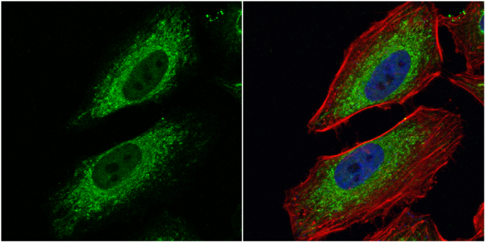
- Pyruvate Dehydrogenase E1 alpha antibody detects Pyruvate Dehydrogenase E1 alpha protein at mitochondria by immunofluorescent analysis.Sample: HeLa cells were fixed in 4% paraformaldehyde at RT for 15 min.Green: Pyruvate Dehydrogenase E1 alpha protein stained by Pyruvate Dehydrogenase E1 alpha antibody (GTX104015) diluted at 1:100.Red: phalloidin, a cytoskeleton marker, stained by phalloidin (invitrogen, A12380) diluted at 1:200.Blue: Hoechst 33342 staining.
Supportive validation
- Submitted by
- GeneTex (provider)
- Main image
- Experimental details
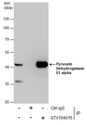
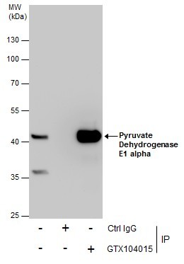
- Immunoprecipitation of Pyruvate Dehydrogenase E1 alpha protein from HepG2 whole cell extracts using 5 £gg of Pyruvate Dehydrogenase E1 alpha antibody (GTX104015).Western blot analysis was performed using Pyruvate Dehydrogenase E1 alpha antibody (GTX104015).EasyBlot anti-Rabbit IgG (GTX221666-01) was used as a secondary reagent.
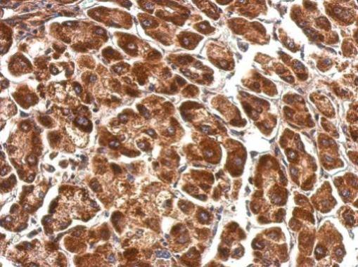
Supportive validation
- Submitted by
- GeneTex (provider)
- Main image

- Experimental details
- Pyruvate Dehydrogenase E1 alpha antibody detects PDHA1 protein at cytosol on human hepatoma by immunohistochemical analysis. Sample: Paraffin-embedded hepatoma. Pyruvate Dehydrogenase E1 alpha antibody (GTX104015) dilution: 1:500.