Explore
 Validate
 Learn
Learn Western blot
 Immunocytochemistry
Antibody data
- Antibody Data
- Antigen structure
- References [1]
- Comments [0]
- Validations
- Immunocytochemistry [2]
- Immunoprecipitation [1]
- Immunohistochemistry [1]
- Other assay [2]
Submit
Validation data
Reference
Comment
Report error
- Product number
- PA5-21536 - Provider product page

- Provider
- Invitrogen Antibodies
- Product name
- PDHA1 Polyclonal Antibody
- Antibody type
- Polyclonal
- Antigen
- Recombinant full-length protein
- Description
- Recommended positive controls: 293T, A431, HeLa, HepG2, Mouse brain, Rat brain. Predicted reactivity: Mouse (98%), Rat (98%), Xenopus laevis (91%), Chicken (90%), Rhesus Monkey (100%), Chimpanzee (100%), Bovine (99%). Store product as a concentrated solution. Centrifuge briefly prior to opening the vial.
- Reactivity
- Human, Mouse, Rat
- Host
- Rabbit
- Isotype
- IgG
- Vial size
- 100 μL
- Concentration
- 1.39 mg/mL
- Storage
- Store at 4°C short term. For long term storage, store at -20°C, avoiding freeze/thaw cycles.
Submitted references Mutations in ARL2BP, a protein required for ciliary microtubule structure, cause syndromic male infertility in humans and mice.
Moye AR, Bedoni N, Cunningham JG, Sanzhaeva U, Tucker ES, Mathers P, Peter VG, Quinodoz M, Paris LP, Coutinho-Santos L, Camacho P, Purcell MG, Winkelmann AC, Foster JA, Pugacheva EN, Rivolta C, Ramamurthy V
PLoS genetics 2019 Aug;15(8):e1008315
PLoS genetics 2019 Aug;15(8):e1008315
No comments: Submit comment
Supportive validation
- Submitted by
- Invitrogen Antibodies (provider)
- Main image

- Experimental details
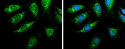
- Immunocytochemistry-Immunofluorescence analysis of PDHA1 was performed in HeLa cells fixed in ice-cold MeOH for 5 min. Green: PDHA1 Polyclonal Antibody (Product # PA5-21536) diluted at 1:2000. Blue: Fluoroshield with DAPI.

- Submitted by
- Invitrogen Antibodies (provider)
- Main image
- Experimental details
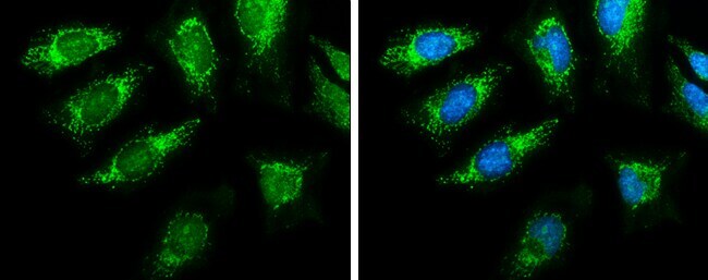
- Immunocytochemistry-Immunofluorescence analysis of PDHA1 was performed in HeLa cells fixed in ice-cold MeOH for 5 min. Green: PDHA1 Polyclonal Antibody (Product # PA5-21536) diluted at 1:2000. Blue: Fluoroshield with DAPI.
Supportive validation
- Submitted by
- Invitrogen Antibodies (provider)
- Main image
- Experimental details
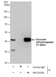
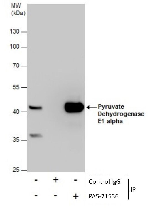
- Immunoprecipitation of Pyruvate Dehydrogenase E1 alpha was performed in HepG2 whole cell extracts using 5 µg of PDHA1 Polyclonal Antibody (Product # PA5-21536). Samples were transferred to a membrane and probed with PDHA1 Polyclonal Antibody as a primary antibody and an HRP-conjugated anti-Rabbit IgG was used as a secondary antibody.
Supportive validation
- Submitted by
- Invitrogen Antibodies (provider)
- Main image
- Experimental details
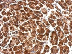
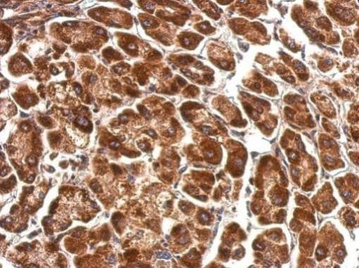
- PDHA1 Polyclonal Antibody detects PDHA1 protein at cytosol on human hepatoma by immunohistochemical analysis. Sample: Paraffin-embedded hepatoma. PDHA1 Polyclonal Antibody (Product # PA5-21536) dilution: 1:500. Antigen Retrieval: EDTA based buffer, pH 8.0, 15 min.
Supportive validation
- Submitted by
- Invitrogen Antibodies (provider)
- Main image

- Experimental details
- Immunoprecipitation of Pyruvate Dehydrogenase E1 alpha was performed in HepG2 whole cell extracts using 5 µg of PDHA1 Polyclonal Antibody (Product # PA5-21536). Samples were transferred to a membrane and probed with PDHA1 Polyclonal Antibody as a primary antibody and an HRP-conjugated anti-Rabbit IgG was used as a secondary antibody.

- Submitted by
- Invitrogen Antibodies (provider)
- Main image
- Experimental details
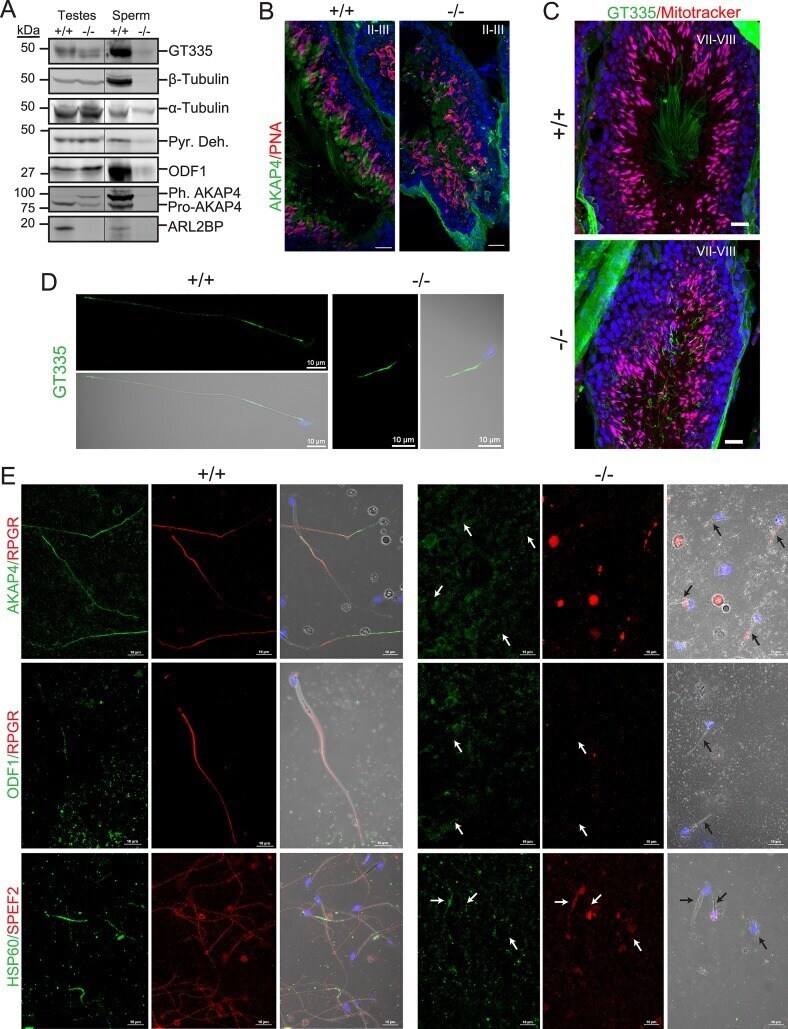
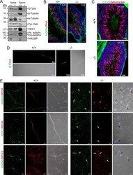
- Fig 6 ARL2BP loss results in impaired sperm tail development. (A) Immunoblot of WT (+/+) and KO (-/-) testis and sperm lysates probed for the indicated sperm tail markers: axonemal markers (Glutamylated tubulin (GT335) and beta-tubulin) and accessory structure markers (Pyruvate dehydrogenase (Pyr. Deh.), Outer Dense Fiber 1 (ODF1), and A-kinase anchoring protein 4 (pre-processed = Pro-AKAP4 and phosphorylated, processed = Ph. AKAP4). Molecular weights in kilodaltons (kDa) are displayed on the left. alpha-tubulin is used as the loading control. (B) WT (+/+) and KO (-/-) murine testes sections stained with PNA lectin (acrosomes, red) and AKAP4 (green), or (C) mitotracker (mitochondria, red) and GT335 (green). The nuclei are stained with DAPI (blue). Scale Bar = 20mum. (D and E) Sperm stained with the indicated sperm tail markers in WT (+/+) and KO (-/-) murine sperm. GT335 -axoneme, green; AKAP4 -fibrous sheath, green; Retinititis Pigmentosa GTPase Regulator (RPGR-axoneme, red); ODF1 -outer dense fibers, green; Heat Shock Protein 60 kDa (HSP60 -mitochondrial sheath, green), and Sperm Flagellar Protein 2 (SPEF2 -axoneme, red). Arrows point to sperm tails. Scale Bar = 10mum.