Explore
 Validate
 Learn
Learn Western blot
 ELISA
ELISAAntibody data
- Antibody Data
- Antigen structure
- References [2]
- Comments [0]
- Validations
- Western blot [1]
Submit
Validation data
Reference
Comment
Report error
- Product number
- A00076-2 - Provider product page

- Provider
- Boster Biological Technology
- Product name
- Anti-LDL Receptor/LDLR Antibody Picoband™
- Antibody type
- Polyclonal
- Description
- Rabbit IgG polyclonal antibody for LDL Receptor/LDLR detection. Tested with WB, IHC-P, FCM, Direct ELISA in Human;Rat.
- Reactivity
- Human, Rat
- Host
- Rabbit
- Vial size
- 100μg/vial
- Concentration
- Add 0.2ml of distilled water will yield a concentration of 500ug/ml.
- Storage
- At -20°C for one year. After reconstitution, at 4°C for one month. It can also be aliquoted and stored frozen at -20°C for a longer time. Avoid repeated freezing and thawing.
- Handling
- Add 0.2ml of distilled water will yield a concentration of 500ug/ml.
Submitted references Difference in LDL receptor feedback regulation in macrophages and vascular smooth muscle cells: foam cell transformation under inflammatory stress.
Inflammatory stress increases unmodified LDL uptake via LDL receptor: an alternative pathway for macrophage foam-cell formation.
Ye Q, Lei H, Fan Z, Zheng W, Zheng S
Inflammation 2014 Apr;37(2):555-65
Inflammation 2014 Apr;37(2):555-65
Inflammatory stress increases unmodified LDL uptake via LDL receptor: an alternative pathway for macrophage foam-cell formation.
Ye Q, Chen Y, Lei H, Liu Q, Moorhead JF, Varghese Z, Ruan XZ
Inflammation research : official journal of the European Histamine Research Society ... [et al.] 2009 Nov;58(11):809-18
Inflammation research : official journal of the European Histamine Research Society ... [et al.] 2009 Nov;58(11):809-18
No comments: Submit comment
Supportive validation
- Submitted by
- Boster Biological Technology (provider)
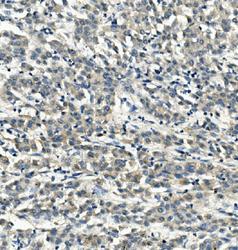
- Main image
- Experimental details
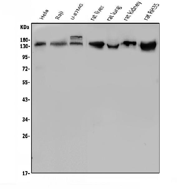
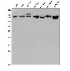
- Western blot analysis of LDLR using anti-LDLR antibody (A00076-2). Electrophoresis was performed on a 5-20% SDS-PAGE gel at 70V (Stacking gel) / 90V (Resolving gel) for 2-3 hours. The sample well of each lane was loaded with 50ug of sample under reducing conditions. Lane 1: human Hela whole cell lysates, Lane 2: human Raji whole cell lysates, Lane 3: human U-87MG whole cell lysates, Lane 4: rat liver tissue lysates, Lane 5: rat lung tissue lysates, Lane 6: rat kidney tissue lysates, Lane 7: rat RH35 whole cell lysates. After Electrophoresis, proteins were transferred to a Nitrocellulose membrane at 150mA for 50-90 minutes. Blocked the membrane with 5% Non-fat Milk/ TBS for 1.5 hour at RT. The membrane was incubated with rabbit anti-LDLR antigen affinity purified polyclonal antibody (Catalog # A00076-2) at 0.5 μg/mL overnight at 4°C, then washed with TBS-0.1%Tween 3 times with 5 minutes each and probed with a goat anti-rabbit IgG-HRP secondary antibody at a dilution of 1:5000 for 1.5 hour at RT. The signal is developed using an Enhanced Chemiluminescent detection (ECL) kit (Catalog # EK1002) with Tanon 5200 system. A specific band was detected for LDLR at approximately 130KD. The expected band size for LDLR is at 95KD.
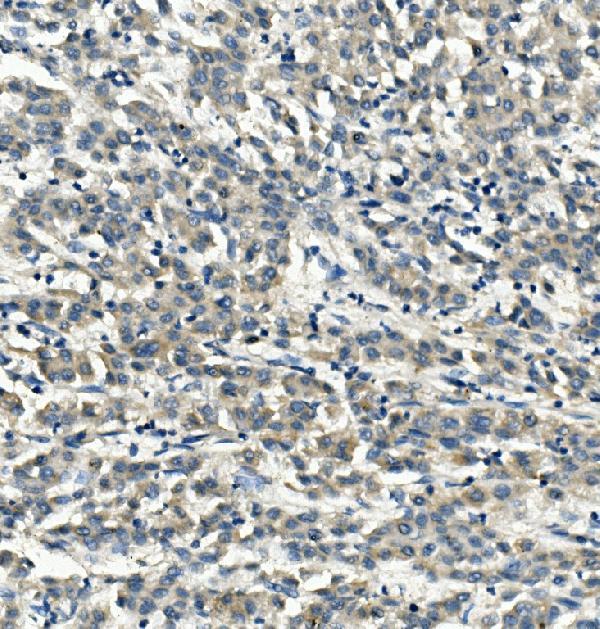
- Additional image