Explore
 Validate
 Learn
Learn Western blot
Antibody data
- Antibody Data
- Antigen structure
- References [4]
- Comments [0]
- Validations
- Western blot [2]
- Immunocytochemistry [1]
- Immunohistochemistry [1]
Submit
Validation data
Reference
Comment
Report error
- Product number
- AF2229 - Provider product page

- Provider
- R&D Systems
- Product name
- Human Nectin-2/CD112 Antibody
- Antibody type
- Polyclonal
- Description
- Antigen Affinity-purified. Detects human Nectin-2/CD112 in direct ELISAs and Western blots. In direct ELISAs and Western blots, less than 2% cross-reactivity with recombinant human (rh) Nectin-1, rhNectin-3 and rhNectin-4 is observed.
- Reactivity
- Human
- Host
- Goat
- Conjugate
- Unconjugated
- Antigen sequence
NP_002847- Isotype
- IgG
- Vial size
- 100 ug
- Concentration
- LYOPH
- Storage
- Use a manual defrost freezer and avoid repeated freeze-thaw cycles. 12 months from date of receipt, -20 to -70 °C as supplied. 1 month, 2 to 8 °C under sterile conditions after reconstitution. 6 months, -20 to -70 °C under sterile conditions after reconstitution.
Submitted references The Ubiquitin-proteasome pathway regulates Nectin2/CD112 expression and impairs NK cell recognition and killing.
Blockade of the checkpoint receptor TIGIT prevents NK cell exhaustion and elicits potent anti-tumor immunity.
Low-dose bortezomib increases the expression of NKG2D and DNAM-1 ligands and enhances induced NK and γδ T cell-mediated lysis in multiple myeloma.
p120 regulates endothelial permeability independently of its NH2 terminus and Rho binding.
Molfetta R, Milito ND, Zitti B, Lecce M, Fionda C, Cippitelli M, Santoni A, Paolini R
European journal of immunology 2019 Jun;49(6):873-883
European journal of immunology 2019 Jun;49(6):873-883
Blockade of the checkpoint receptor TIGIT prevents NK cell exhaustion and elicits potent anti-tumor immunity.
Zhang Q, Bi J, Zheng X, Chen Y, Wang H, Wu W, Wang Z, Wu Q, Peng H, Wei H, Sun R, Tian Z
Nature immunology 2018 Jul;19(7):723-732
Nature immunology 2018 Jul;19(7):723-732
Low-dose bortezomib increases the expression of NKG2D and DNAM-1 ligands and enhances induced NK and γδ T cell-mediated lysis in multiple myeloma.
Niu C, Jin H, Li M, Zhu S, Zhou L, Jin F, Zhou Y, Xu D, Xu J, Zhao L, Hao S, Li W, Cui J
Oncotarget 2017 Jan 24;8(4):5954-5964
Oncotarget 2017 Jan 24;8(4):5954-5964
p120 regulates endothelial permeability independently of its NH2 terminus and Rho binding.
Herron CR, Lowery AM, Hollister PR, Reynolds AB, Vincent PA
American journal of physiology. Heart and circulatory physiology 2011 Jan;300(1):H36-48
American journal of physiology. Heart and circulatory physiology 2011 Jan;300(1):H36-48
No comments: Submit comment
Supportive validation
- Submitted by
- R&D Systems (provider)
- Main image
- Experimental details
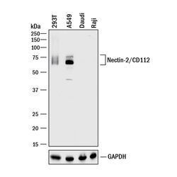
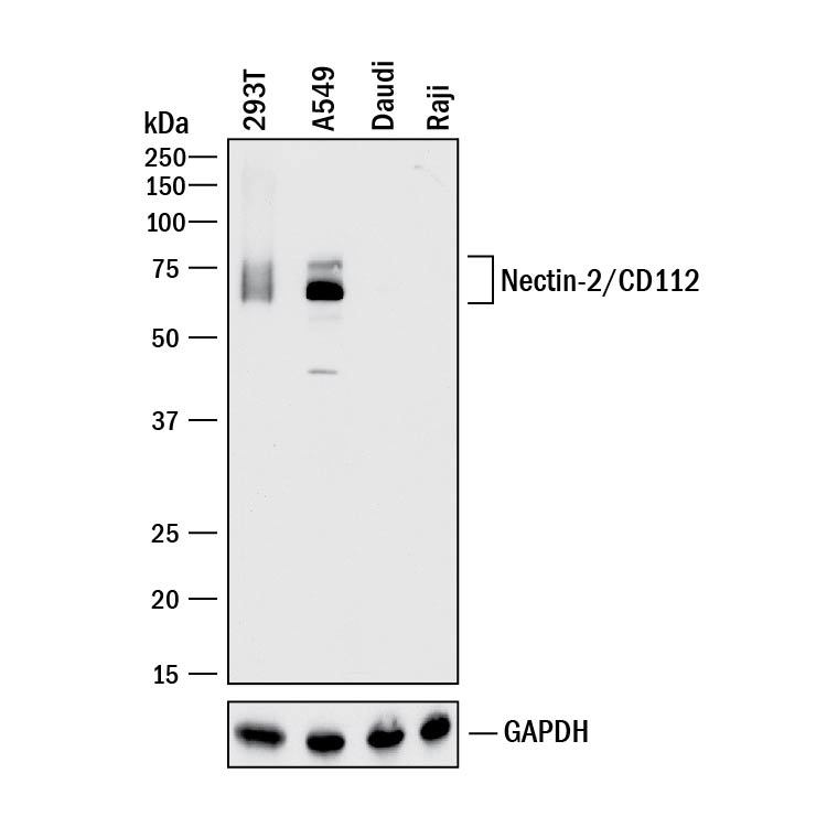
- Detection of Human Nectin-2/CD112 by Western Blot. Western blot shows lysates of 293T human embryonic kidney cell line, A549 human lung carcinoma cell line, Daudi human Burkitt's lymphoma cell line, and Raji human Burkitt's lymphoma cell line. PVDF membrane was probed with 0.1 µg/mL of Goat Anti-Human Nectin-2/CD112 Antigen Affinity-purified Polyclonal Antibody (Catalog # AF2229) followed by HRP-conjugated Anti-Goat IgG Secondary Antibody (Catalog # HAF017). A specific band was detected for Nectin-2/CD112 at approximately 60-75 kDa (as indicated). Daudi human Burkitt's lymphoma cell line and Raji human Burkitt's lymphoma cell line are shown as negative controls. GAPDH (Catalog # AF5718) is shown as a loading control.This experiment was conducted under reducing conditions and using Immunoblot Buffer Group 1.
- Submitted by
- R&D Systems (provider)
- Main image

- Experimental details
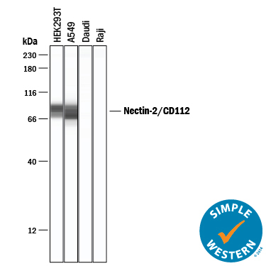
- Detection of Human Nectin-2/CD112 by Simple WesternTM. Simple Western lane view shows lysates of HEK293T human embryonic kidney cell line, A549 human lung carcinoma cell line, Daudi human Burkitt's lymphoma cell line, and Raji human Burkitt's lymphoma cell line, loaded at 0.2 mg/mL. A specific band was detected for Nectin-2/CD112 at approximately 71-86 kDa (as indicated) using 20 µg/mL of Goat Anti-Human Nectin-2/CD112 Antigen Affinity-purified Polyclonal Antibody (Catalog # AF2229) followed by 1:50 dilution of HRP-conjugated Anti-Goat IgG Secondary Antibody (Catalog # HAF109). This experiment was conducted under reducing conditions and using the 12-230 kDa separation system.
Supportive validation
- Submitted by
- R&D Systems (provider)
- Main image

- Experimental details
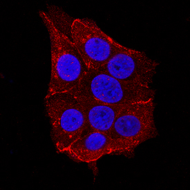
- Nectin-2/CD112 in MCF-7 Human Cell Line. Nectin-2/CD112 was detected in immersion fixed MCF-7 human breast cancer cell line using Goat Anti-Human Nectin-2/CD112 Antigen Affinity-purified Polyclonal Antibody (Catalog # AF2229) at 1.7 µg/mL for 3 hours at room temperature. Cells were stained using the NorthernLights™ 557-conjugated Anti-Goat IgG Secondary Antibody (red; Catalog # NL001) and counterstained with DAPI (blue). Specific staining was localized to cytoplasm. View our protocol for Fluorescent ICC Staining of Cells on Coverslips.
Supportive validation
- Submitted by
- R&D Systems (provider)
- Main image
- Experimental details
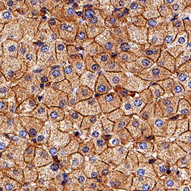
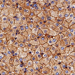
- Nectin-2/CD112 in Human Liver. Nectin-2/CD112 was detected in immersion fixed paraffin-embedded sections of human liver using Goat Anti-Human Nectin-2/CD112 Antigen Affinity-purified Polyclonal Antibody (Catalog # AF2229) at 3 µg/mL for 1 hour at room temperature followed by incubation with the Anti-Goat IgG VisUCyte™ HRP Polymer Antibody (Catalog # VC004). Tissue was stained using DAB (brown) and counterstained with hematoxylin (blue). Specific staining was localized to cell membrane. View our protocol for IHC Staining with VisUCyte HRP Polymer Detection Reagents.