Explore
 Validate
 Learn
Learn Western blot
 Immunoprecipitation
Antibody data
- Antibody Data
- Antigen structure
- References [0]
- Comments [0]
- Validations
- Western blot [2]
- Immunocytochemistry [1]
- Chromatin Immunoprecipitation [2]
Submit
Validation data
Reference
Comment
Report error
- Product number
- ABIN2616351 - Provider product page

- Provider
- antibodies-online
- Product name
- anti-Ankyrin Repeat, Family A (RFXANK-Like), 2 (ANKRA2) (AA 1-313) antibody
- Antibody type
- Polyclonal
- Description
- Antiserum
- Reactivity
- Human, Mouse
- Host
- Rabbit
- Epitope
- AA 1-313
- Isotype
- IgG
- Vial size
- 100 μL
- Storage
- May be stored at 4°C for short-term only. Aliquot to avoid freeze-thaw cycles. Store at -20°C. Aliquots are stable for at least 1 year.
- Handling
- Aliquot to avoid repeated freezing and thawing.
No comments: Submit comment
Supportive validation
- Submitted by
- antibodies-online (provider)
- Main image
- Experimental details
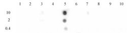
- Histone H3 acetyl Lys9 antibody tested by dot blot analysis. Dot blot analysis was used to confirm the specificity of Histone H3 acetyl Lys9 antibody for acetyl Lys9 histone H3. Acetylated peptides corresponding to the immunogen and related peptides were spotted onto PVDF and probed with Histone H3 acetyl Lys9 antibody at a dilution of 1 μg/ml. The amount of peptide (picomoles) spotted is indicated next to each row. Lane 1: histone H3 acetyl-Lys4 peptide. Lane 2: unmodified Lys4 peptide. Lane 3: acetyl-Lys18 peptide. Lane 4: unmodified Lys9/14/18 peptide. Lane 5: acetyl-Lys9 peptide. Lane 6: acetyl- Lys14 peptide. Lane 7: acetyl-Lys18 peptide. Lane 8: acetyl-Lys23 peptide. Lane 9: acetyl-Lys27 peptide. Lane 10: unmodified Lys27 peptide.

- Submitted by
- antibodies-online (provider)
- Main image
- Experimental details
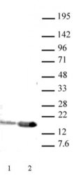
- Western blot of Histone H3 acetyl Lys9 antibody (pAb). HeLa nuclear extract (20 μg per lane) probed with Histone H3 acetyl Lys9 antibody (1 μg per ml). Lane 1: no treatment. Lane 2: cells treated with sodium butyrate.

Supportive validation
- Submitted by
- antibodies-online (provider)
- Main image
- Experimental details
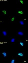
- Immunofluorescence stain of Histone H3 acetyl Lys9 pAb. HeLa cells stained at 2 μg/ml with Histone H3 acetyl Lys9 antibody. Top panel: Histone H3 acetyl Lys9 antibody. Middle panel: DAPI. Bottom panel: merge.

Supportive validation
- Submitted by
- antibodies-online (provider)
- Main image
- Experimental details
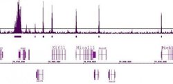
- Histone H3 Acetyl Lys9 antibody (pAb) tested by ChIP-Seq. ChIP was performed using the ChIP-IT® High Sensitivity Kit (Cat. No. 53040) with 30 ug of chromatin from mouse liver. ChIP DNA was sequenced on the Illumina GA II and 25 million sequence tags were mapped to identify H3K9Ac binding across the genome. The image shows a 1.5 million base pair region on chromosome 15. H3K9Ac shows promoter localization at many genes and broader binding near the Gcat gene.

- Submitted by
- antibodies-online (provider)
- Main image

- Experimental details
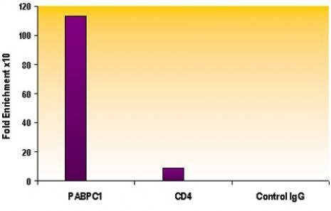
- ChIP of Histone H3 acetyl Lys9 pAb. Chromatin IP performed using the ChIP-IT® Express Kit (Catalog No. 53008) and HeLa Chromatin (1.5 x 10⁶ cell equivalents per ChIP) using 3 μg of Histone H3 acetyl Lys9 antibody or the equivalent amount of rabbit IgG as a negative control. Real time, quantitative PCR (RT-qPCR) was performed on DNA purified from each of the ChIP reactions using a primer pair specific for the indicated gene. Data are presented as Fold Enrichment of the ChIP antibody signal versus the negative control IgG using the ddCT method.