Explore
 Validate
 Learn
Learn Western blot
 Immunohistochemistry
Antibody data
- Antibody Data
- Antigen structure
- References [2]
- Comments [0]
- Validations
- Immunohistochemistry [1]
Submit
Validation data
Reference
Comment
Report error
- Product number
- HPA029564 - Provider product page

- Provider
- Atlas Antibodies
- Proper citation
- Atlas Antibodies Cat#HPA029564, RRID:AB_10602213
- Product name
- Anti-CYP2E1
- Antibody type
- Polyclonal
- Description
- Polyclonal Antibody against Human CYP2E1, Gene description: cytochrome P450, family 2, subfamily E, polypeptide 1, Alternative Gene Names: CYP2E, Validated applications: WB, IHC, Uniprot ID: P05181, Storage: Store at +4°C for short term storage. Long time storage is recommended at -20°C.
- Reactivity
- Human
- Host
- Rabbit
- Conjugate
- Unconjugated
- Isotype
- IgG
- Vial size
- 100 µl
- Concentration
- 0.1 mg/ml
- Storage
- Store at +4°C for short term storage. Long time storage is recommended at -20°C.
- Handling
- The antibody solution should be gently mixed before use.
Submitted references Analysis of the Human Tissue-specific Expression by Genome-wide Integration of Transcriptomics and Antibody-based Proteomics
Analysis of the Human Tissue-specific Expression by Genome-wide Integration of Transcriptomics and Antibody-based Proteomics
Fagerberg L, Hallström B, Oksvold P, Kampf C, Djureinovic D, Odeberg J, Habuka M, Tahmasebpoor S, Danielsson A, Edlund K, Asplund A, Sjöstedt E, Lundberg E, Szigyarto C, Skogs M, Takanen J, Berling H, Tegel H, Mulder J, Nilsson P, Schwenk J, Lindskog C, Danielsson F, Mardinoglu A, Sivertsson Å, von Feilitzen K, Forsberg M, Zwahlen M, Olsson I, Navani S, Huss M, Nielsen J, Ponten F, Uhlén M
Molecular & Cellular Proteomics 2014;13(2):397-406
Molecular & Cellular Proteomics 2014;13(2):397-406
Analysis of the Human Tissue-specific Expression by Genome-wide Integration of Transcriptomics and Antibody-based Proteomics
Fagerberg L, Hallstrom B, Oksvold P, Kampf C, Djureinovic D, Odeberg J, Habuka M, Tahmasebpoor S, Danielsson A, Edlund K, Asplund A, Sjostedt E, Lundberg E, Szigyarto C, Skogs M, Takanen J, Berling H, Tegel H, Mulder J, Nilsson P, Schwenk J, Lindskog C, Danielsson F, Mardinoglu A, Sivertsson A, von Feilitzen K, Forsberg M, Zwahlen M, Olsson I, Navani S, Huss M, Nielsen J, Ponten F, Uhlen M
Molecular & Cellular Proteomics 2014 January;13(2):397-406
Molecular & Cellular Proteomics 2014 January;13(2):397-406
No comments: Submit comment
Supportive validation
- Submitted by
- Atlas Antibodies (provider)
- Enhanced method
- Orthogonal validation
- Main image
- Experimental details
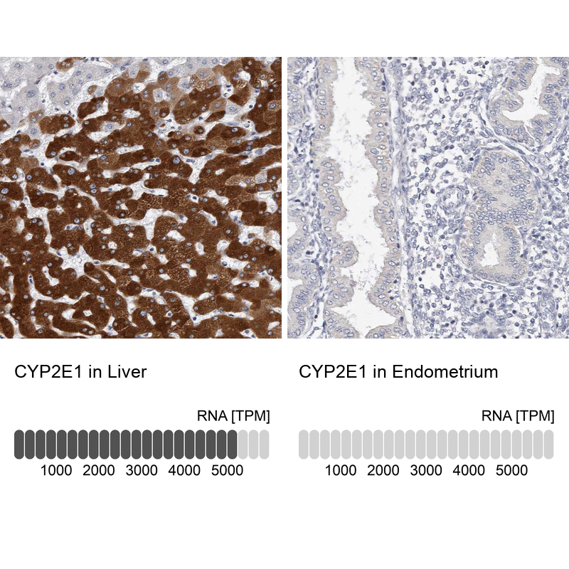
- Immunohistochemistry analysis in human liver and endometrium tissues using HPA029564 antibody. Corresponding CYP2E1 RNA-seq data are presented for the same tissues.
- Sample type
- Human
- Protocol
- Protocol