Explore
 Validate
 Learn
Learn Western blot
 Immunocytochemistry
 Chromatin Immunoprecipitation
Antibody data
- Antibody Data
- Antigen structure
- References [3]
- Comments [0]
- Validations
- Immunocytochemistry [1]
- Immunohistochemistry [1]
- Other assay [3]
Submit
Validation data
Reference
Comment
Report error
- Product number
- PA5-23065 - Provider product page

- Provider
- Invitrogen Antibodies
- Product name
- JMJD2C Polyclonal Antibody
- Antibody type
- Polyclonal
- Antigen
- Recombinant full-length protein
- Description
- Prior to immunostaining paraffin tissues, antigen retrieval with sodium citrate buffer (pH 6.0) is recommended.
- Reactivity
- Human, Mouse
- Host
- Rabbit
- Isotype
- IgG
- Vial size
- 100 μL
- Concentration
- 1.0 mg/mL
- Storage
- Store at 4°C short term. For long term storage, store at -20°C, avoiding freeze/thaw cycles.
Submitted references RNA-seq analysis of PHD and VHL inhibitors reveals differences and similarities to the hypoxia response.
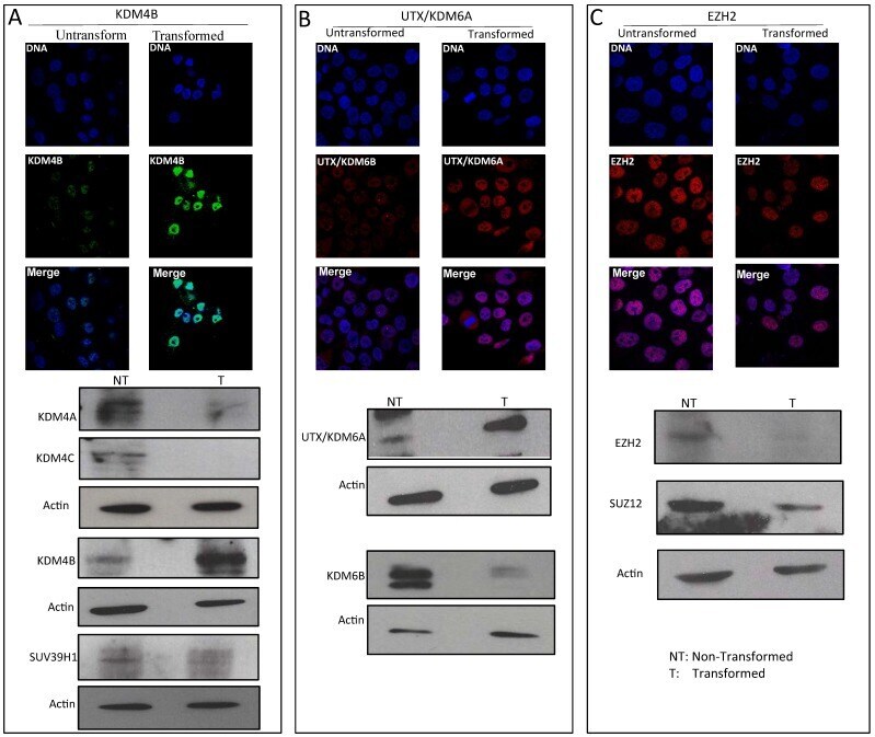
Heterochromatin Reduction Correlates with the Increase of the KDM4B and KDM6A Demethylases and the Expression of Pericentromeric DNA during the Acquisition of a Transformed Phenotype.
PITX1, a specificity determinant in the HIF-1α-mediated transcriptional response to hypoxia.
Frost J, Ciulli A, Rocha S
Wellcome open research 2019;4:17
Wellcome open research 2019;4:17
Heterochromatin Reduction Correlates with the Increase of the KDM4B and KDM6A Demethylases and the Expression of Pericentromeric DNA during the Acquisition of a Transformed Phenotype.
Gurrion C, Uriostegui M, Zurita M
Journal of Cancer 2017;8(14):2866-2875
Journal of Cancer 2017;8(14):2866-2875
PITX1, a specificity determinant in the HIF-1α-mediated transcriptional response to hypoxia.
Mudie S, Bandarra D, Batie M, Biddlestone J, Moniz S, Ortmann B, Shmakova A, Rocha S
Cell cycle (Georgetown, Tex.) 2014;13(24):3878-91
Cell cycle (Georgetown, Tex.) 2014;13(24):3878-91
No comments: Submit comment
Supportive validation
- Submitted by
- Invitrogen Antibodies (provider)
- Main image
- Experimental details
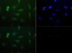
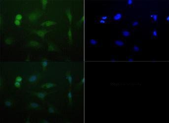
- Immunofluorescent analysis of JMJD2C using a polyclonal antibody (Product # PA5-23065).
Supportive validation
- Submitted by
- Invitrogen Antibodies (provider)
- Main image
- Experimental details
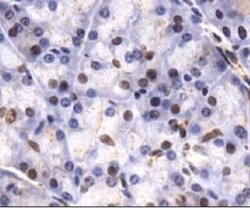
- Immunohistochemical analysis of JMJD2C in paraffin embedded mouse pancreas. Samples were incubated in JMJD2C polyclonal antibody (Product # PA5-23065) followed by DAB with hematoxylin counterstain.

Supportive validation
- Submitted by
- Invitrogen Antibodies (provider)
- Main image
- Experimental details
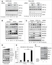
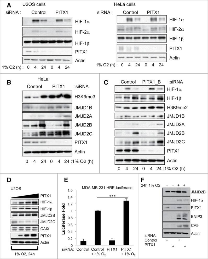
- Figure 2. PITX1 is a specificity determinant for HIF-1a-dependent target gene activation. (A) U2OS and HeLa cells were transfected with control or PITX1 siRNA, prior to treatment with 1% O2 for 24 hours. Whole cell lysates were obtained 48 hours post-transfection and analyzed by protein gel blot using the indicated antibodies. (B) HeLa cells were transfected with control or PITX1 siRNA, prior to treatment with 1% O2 for the indicated periods of time. Whole cell lysates were obtained 48 hours post-transfection and analyzed by western blot using the indicated antibodies. (C) HeLa cells were transfected, processed and analyzed as in B. (D) U2OS were co-transfected with 1 mug of GFP-HIF-1beta and increasing amounts of PITX1 plasmid (0.1, 0.25 and 0.5 mug) prior to exposure to 1% O2 for 24 hours. Whole cell lysates were obtained 48 hours post-transfection and analyzed by protein gel blot using the indicated antibodies. (E) MDA-MB-231-HRE cells were transfected with control and PITX1 siRNA oligonucleotides prior to treatment with 1% O2 for 24 hours. Luciferase activity was measured 48 hours post-transfection. Graph depicts mean and standard deviation of a minimum of 3 independent experiments. Student's t-test was performed to calculate p values, and levels of significance are denoted as follows: * P < 0.05, ** P < 0.01, and *** P < 0.001. (F) MDA-MB-231 cells were transfected with control and PITX1 siRNA oligonucleotides prior to treatment to 1% O2 for 24 hours. Whole cell lys
- Submitted by
- Invitrogen Antibodies (provider)
- Main image
- Experimental details
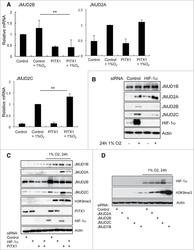
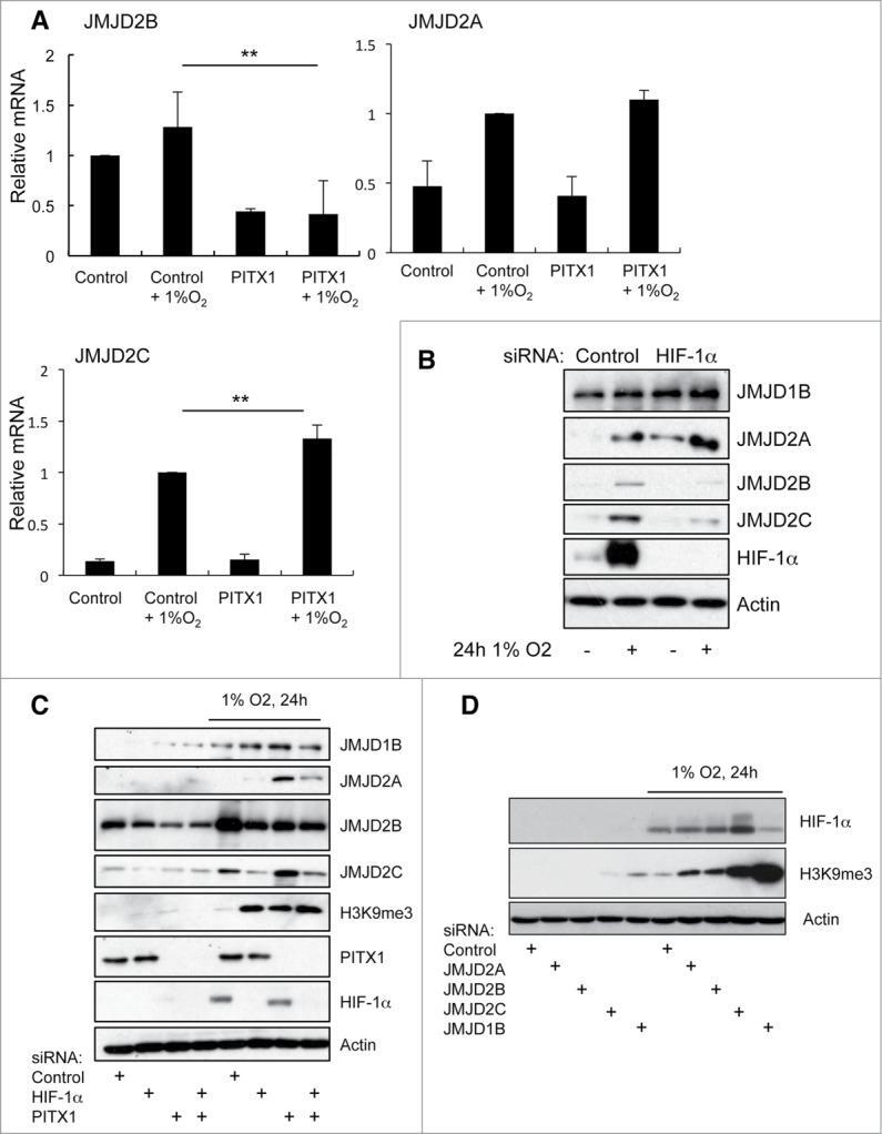
- Figure 3. PITX1 regulates JMJD2B and JMJD2C expression in a HIF-1alpha-dependent manner. (A) HeLa cells were transfected with control or PITX1 siRNA, prior to treatment with 1% O2 for 24 hours. Total mRNA was extracted and levels of JMJD2A, JMJD2B and JMJD2C mRNA were analyzed by qPCR. Graph depicts mean and standard deviation of a minimum of 3 independent experiments. Data was normalized using actin and compared to control siRNA. Student's t-test was performed to calculate p values, and levels of significance are denoted as follows: * P < 0.05, ** P < 0.01, and *** P < 0.001. (B) HeLa cells were transfected with control or HIF-1alpha siRNA, prior to treatment with 1% O2 for 24 hours. Whole cell lysates were obtained 48 hours post-transfection and analyzed by protein gel blot using the indicated antibodies. (C) HeLa cells were transfected with control, PITX1, HIF-1a or PITX1 and HIF-1alpha siRNA, prior to treatment with 1% O2 for 24 hours. Whole cell lysates were obtained 48 hours post-transfection and analyzed by western blot using the indicated antibodies. (D) HeLa cells were transfected with the indicated siRNAs oligonucleotides prior to treatment with 1% O2 for 24 hours. Whole cell lysates were obtained 48 hours post-transfection and analyzed by protein gel blot using the indicated antibodies.
- Submitted by
- Invitrogen Antibodies (provider)
- Main image
- Experimental details
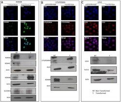
- Figure 3 Variation in the levels of enzymes that modify histone H3 in transformed MCF10A-Er-Src cell line . A, Immunostaining to detect KDM4B demethylase and western blots detecting KDM4A, KDM4B and KDM4C histone demethylases as well as the methyl transferase SUV39H1 levels in non-transformed and transformed cells. B, Immunostaining to detect UTX/KDM6A and western blot experiments detecting UTX/KDM6A and KDM6B in non-transformed and transformed cells. C, Immunostaining to detect the methyl transferase EZH2 and western blots to detect EZH2 and SUZ12 in transformed and no-transformed cells. Acting was used as loading control in all the western blot experiments. NT=Non-transformed cells; T= Transformed cells.