Explore
 Validate
 Learn
Learn Western blot
 ELISA
ELISAAntibody data
- Antibody Data
- Antigen structure
- References [2]
- Comments [0]
- Validations
- Western blot [1]
- Immunocytochemistry [1]
- Immunohistochemistry [1]
- Flow cytometry [1]
Submit
Validation data
Reference
Comment
Report error
- Product number
- ABIN954029 - Provider product page

- Provider
- antibodies-online
- Product name
- anti-Protocadherin 20 (PCDH20) (AA 452-480), (Middle Region) antibody
- Antibody type
- Polyclonal
- Antigen
- KLH conjugated synthetic peptide between 452-480 amino acids from the Central region of human PCDH20
- Description
- Affinity chromatography on Protein A
- Reactivity
- Human
- Host
- Rabbit
- Epitope
- AA 452-480,Middle Region
- Vial size
- 0.4 mL
- Concentration
- 0.25 mg/mL
- Storage
- Store the antibody undiluted at 2-8°C for one month or (in aliquots) at -20°C for longer.
- Handling
- Avoid repeated freezing and thawing.
Submitted references Recent progress in protocadherin research.
Phylogenetic analysis of the cadherin superfamily allows identification of six major subfamilies besides several solitary members.
Suzuki ST
Experimental cell research 2000 Nov 25;261(1):13-8
Experimental cell research 2000 Nov 25;261(1):13-8
Phylogenetic analysis of the cadherin superfamily allows identification of six major subfamilies besides several solitary members.
Nollet F, Kools P, van Roy F
Journal of molecular biology 2000 Jun 9;299(3):551-72
Journal of molecular biology 2000 Jun 9;299(3):551-72
No comments: Submit comment
Supportive validation
- Submitted by
- antibodies-online (provider)
- Main image
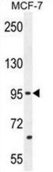
- Experimental details
- PCDH20 Antibody (Center) (Cat. #AP53195PU-N) western blot analysis in MCF-7 cell line lysates (35μg/lane).This demonstrates the PCDH20 antibody detected the PCDH20 protein (arrow).

Supportive validation
- Submitted by
- antibodies-online (provider)
- Main image
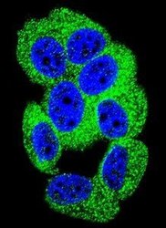
- Experimental details
- Confocal immunofluorescent analysis of PCDH20 Antibody (Center)(Cat#AP53195PU-N) with MCF-7 cell followed by Alexa Fluor 488-conjugated goat anti-rabbit lgG (green).DAPI was used to stain the cell nuclear (blue).

Supportive validation
- Submitted by
- antibodies-online (provider)
- Main image
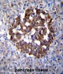
- Experimental details
- PCDH20 Antibody (Center) (Cat. #AP53195PU-N)immunohistochemistry analysis in formalin fixed and paraffin embedded human pancreas tissue followed by peroxidase conjugation of the secondary antibody and DAB staining.This data demonstrates the use of PCDH20 Antibody (Center) for immunohistochemistry. Clinical relevance has not been evaluated.

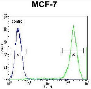
Supportive validation
- Submitted by
- antibodies-online (provider)
- Main image
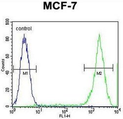
- Experimental details
- PCDH20 Antibody (Center) (Cat. #AP53195PU-N) flow cytometric analysis of MCF-7 cells (right histogram) compared to a negative control cell (left histogram).FITC-conjugated goat-anti-rabbit secondary antibodies were used for the analysis.