Explore
 Validate
 Learn
Learn Western blot
 Immunohistochemistry
Antibody data
- Antibody Data
- Antigen structure
- References [3]
- Comments [0]
- Validations
- Immunohistochemistry [3]
- Flow cytometry [2]
- Other assay [1]
Submit
Validation data
Reference
Comment
Report error
- Product number
- PA1-16635 - Provider product page

- Provider
- Invitrogen Antibodies
- Product name
- LYVE1 Polyclonal Antibody
- Antibody type
- Polyclonal
- Antigen
- Synthetic peptide
- Description
- Prior to immunostaining paraffin tissues, antigen retrieval with sodium citrate buffer (pH 6.0) is recommended. Suggested positive control: mouse spleen lysate or protein.
- Reactivity
- Human, Mouse, Rat
- Host
- Rabbit
- Isotype
- IgG
- Vial size
- 100 μL
- Concentration
- 1 mg/mL
- Storage
- Store at 4°C short term. For long term storage, store at -20°C, avoiding freeze/thaw cycles.
Submitted references Pilot study supporting the existence of novel lymphatic channels within the canine anterior uveal tract using Lyve-1 and CD31.
Systematic Evaluation of Chemically Distinct Tissue Optical Clearing Techniques in Murine Lymph Nodes.
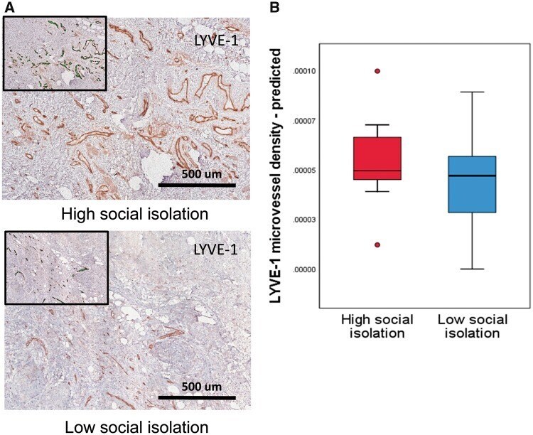
Prometastatic Molecular Profiles in Breast Tumors From Socially Isolated Women.
Dubin A, Freeman KS, Charles J, Ammar DA, Ehrhart EJ
Veterinary ophthalmology 2021 Jul;24(4):354-360
Veterinary ophthalmology 2021 Jul;24(4):354-360
Systematic Evaluation of Chemically Distinct Tissue Optical Clearing Techniques in Murine Lymph Nodes.
Matryba P, Sosnowska A, Wolny A, Bozycki L, Greig A, Grzybowski J, Stefaniuk M, Nowis D, Gołąb J
Journal of immunology (Baltimore, Md. : 1950) 2020 Mar 1;204(5):1395-1407
Journal of immunology (Baltimore, Md. : 1950) 2020 Mar 1;204(5):1395-1407
Prometastatic Molecular Profiles in Breast Tumors From Socially Isolated Women.
Bower JE, Shiao SL, Sullivan P, Lamkin DM, Atienza R, Mercado F, Arevalo J, Asher A, Ganz PA, Cole SW
JNCI cancer spectrum 2018 Jul;2(3):pky029
JNCI cancer spectrum 2018 Jul;2(3):pky029
No comments: Submit comment
Supportive validation
- Submitted by
- Invitrogen Antibodies (provider)
- Main image
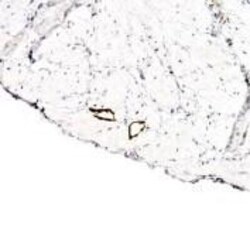
- Experimental details
- Immunohistochemical analysis of LYVE1 in endothelial cells of human bladder vasculature. Samples were incubated in LYVE1 polyclonal antibody (Product # PA1-16635).

- Submitted by
- Invitrogen Antibodies (provider)
- Main image
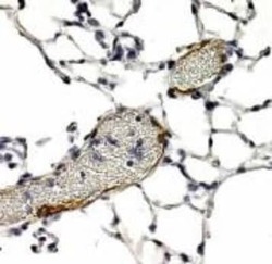
- Experimental details
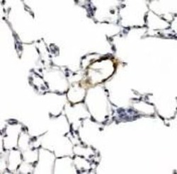
- Immunohistochemical analysis of LYVE1 in endothelial cells of human lung blood vessels. Samples were incubated in LYVE1 polyclonal antibody (Product # PA1-16635). Note presence of RBCs within vessel lumen.
- Submitted by
- Invitrogen Antibodies (provider)
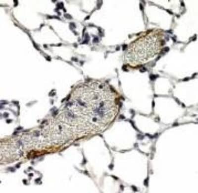
- Main image
- Experimental details
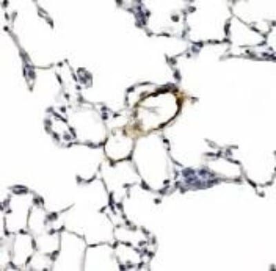
- Immunohistochemical analysis of LYVE1 in endothelial cells of human lung blood vessels. Samples were incubated in LYVE1 polyclonal antibody (Product # PA1-16635).
Supportive validation
- Submitted by
- Invitrogen Antibodies (provider)
- Main image
- Experimental details
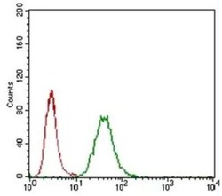
- Flow cytometry of LYVE1 in A549 cells. Samples were incubated in LYVE1 polyclonal antibody (Product # PA1-16635) using a dilution of 1:400 followed by a Alexa Fluor 488 secondary (shown in green). Unstained cells (shown in red).

- Submitted by
- Invitrogen Antibodies (provider)
- Main image

- Experimental details
- Flow cytometry of LYVE1 in A549 cells. Samples were incubated in LYVE1 polyclonal antibody (Product # PA1-16635) using a dilution of 1:400 followed by a Alexa Fluor 488 secondary (shown in green). Unstained cells (shown in red).

Supportive validation
- Submitted by
- Invitrogen Antibodies (provider)
- Main image

- Experimental details
- Figure 2. Analyses of lymphovascular invasion in primary breast tumors indicated increased intratumoral lymphatic vessel endothelial hyaluronan receptor 1 (LYVE-1) microvessel density in tumors from socially isolated women. A) Representative images of LYVE-1-stained sections from patients reporting high vs low social isolation. B) Box plots of predicted scores for LYVE-1 microvessel density in the high- and low-social isolation groups. Each box indicates the interquartile range, and the middle line indicates the median for that group. LYVE-1 = lymphatic vessel endothelial hyaluronan receptor 1.