Explore
 Validate
 Learn
Learn Western blot
 ELISA
ELISAAntibody data
- Antibody Data
- Antigen structure
- References [2]
- Comments [0]
- Validations
- Western blot [1]
- Immunocytochemistry [2]
- Immunohistochemistry [2]
Submit
Validation data
Reference
Comment
Report error
- Product number
- DP3513S - Provider product page

- Provider
- Acris Antibodies GmbH
- Proper citation
- Acris Antibodies GmbH Cat#DP3513S, RRID:AB_1004773
- Product name
- anti LYVE-1
- Antibody type
- Polyclonal
- Antigen
- Highly pure recombinant Mouse soluble LYVE-1 (Ala24-Gly228) produced in insect cells (Cat.-No DA3524).This recombinant soluble LYVE-1 consists of amino acid 24 (Ala) to 228 (Gly) and is fused to a C-terminal His-tag (6xHis).
- Reactivity
- Mouse
- Host
- Rabbit
- Vial size
- 0.1 mg
Submitted references Endogenous angiogenesis inhibitor vasohibin1 exhibits broad-spectrum antilymphangiogenic activity and suppresses lymph node metastasis.
Multipotent mesenchymal stem cells acquire a lymphendothelial phenotype and enhance lymphatic regeneration in vivo.
Heishi T, Hosaka T, Suzuki Y, Miyashita H, Oike Y, Takahashi T, Nakamura T, Arioka S, Mitsuda Y, Takakura T, Hojo K, Matsumoto M, Yamauchi C, Ohta H, Sonoda H, Sato Y
The American journal of pathology 2010 Apr;176(4):1950-8
The American journal of pathology 2010 Apr;176(4):1950-8
Multipotent mesenchymal stem cells acquire a lymphendothelial phenotype and enhance lymphatic regeneration in vivo.
Conrad C, Niess H, Huss R, Huber S, von Luettichau I, Nelson PJ, Ott HC, Jauch KW, Bruns CJ
Circulation 2009 Jan 20;119(2):281-9
Circulation 2009 Jan 20;119(2):281-9
No comments: Submit comment
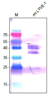
Supportive validation
- Submitted by
- Acris Antibodies GmbH (provider)
- Main image
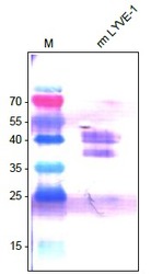
- Experimental details
- Figure 5. Western Analysis of anti-Mouse LYVE-1 Antibody. Sample was loaded in 15% SDS-polyacrylamide gel under reducing conditions.
Supportive validation
- Submitted by
- Acris Antibodies GmbH (provider)
- Main image

- Experimental details
- Figure 1. Staining of mouse colon using a CD31 antibody (green) and Lyve-1 antibody (red). Picturted originate from Dr. Ulrike Fiedler and Stefanie Koidel, Dept. of Vascular Biology and Angiogenesis Research Tumor Biology center, Freiburg, Germany.

- Submitted by
- Acris Antibodies GmbH (provider)
- Main image
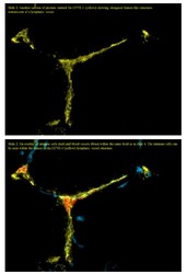
- Experimental details
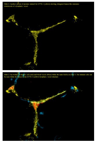
- Figure 4. LYVE-1 and CD31 staining on frozen sections (5μm) of the Mouse prostate.The experiments were performed by Scott Gerber & Edith Lord, PhD, University of Rochester, USA.
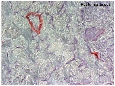
Supportive validation
- Submitted by
- Acris Antibodies GmbH (provider)
- Main image
- Experimental details
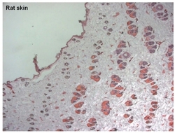
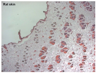
- Figure 2. Immunohistochemistry with Cryo sections from Rat Skin using anti-Mouse LYVE-1 antibody (DP3513/DP3513S)
- Submitted by
- Acris Antibodies GmbH (provider)
- Main image
- Experimental details
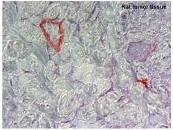
- Figure 3. Immunohistochemistry with Cryo sections from Rat Tumor Tissue using anti-Mouse LYVE-1 antibody (DP3513/DP3513S)