Explore
 Validate
 Learn
Learn Western blot
 Immunocytochemistry
Antibody data
- Antibody Data
- Antigen structure
- References [4]
- Comments [0]
- Validations
- Western blot [2]
- Immunohistochemistry [4]
- Flow cytometry [5]
Submit
Validation data
Reference
Comment
Report error
- Product number
- NB100-725 - Provider product page

- Provider
- Novus Biologicals
- Proper citation
- Novus Cat#NB100-725, RRID:AB_10003273
- Product name
- Rabbit Polyclonal LYVE-1 Antibody
- Antibody type
- Polyclonal
- Description
- Immunogen affinity purified.
- Reactivity
- Human, Mouse, Rat
- Host
- Rabbit
- Isotype
- IgG
- Vial size
- 0.1 ml
- Concentration
- 1.0 mg/ml
- Storage
- Store at 4C short term. Aliquot and store at -20C long term. Avoid freeze-thaw cycles.
Submitted references Lymphatic vessels in human adipose tissue.
Brain-to-cervical lymph node signaling after stroke.
A Glial Signature and Wnt7 Signaling Regulate Glioma-Vascular Interactions and Tumor Microenvironment.
Trafficking of a dual-modality magnetic resonance and fluorescence imaging superparamagnetic iron oxide-based nanoprobe to lymph nodes.
Redondo PAG, Gubert F, Zaverucha-do-Valle C, Dutra TPP, Ayres-Silva JP, Fernandes N, de Souza AAP, Loizidou M, Takiya CM, Rossi MID, Borojevic R
Cell and tissue research 2020 Mar;379(3):511-520
Cell and tissue research 2020 Mar;379(3):511-520
Brain-to-cervical lymph node signaling after stroke.
Esposito E, Ahn BJ, Shi J, Nakamura Y, Park JH, Mandeville ET, Yu Z, Chan SJ, Desai R, Hayakawa A, Ji X, Lo EH, Hayakawa K
Nature communications 2019 Nov 22;10(1):5306
Nature communications 2019 Nov 22;10(1):5306
A Glial Signature and Wnt7 Signaling Regulate Glioma-Vascular Interactions and Tumor Microenvironment.
Griveau A, Seano G, Shelton SJ, Kupp R, Jahangiri A, Obernier K, Krishnan S, Lindberg OR, Yuen TJ, Tien AC, Sabo JK, Wang N, Chen I, Kloepper J, Larrouquere L, Ghosh M, Tirosh I, Huillard E, Alvarez-Buylla A, Oldham MC, Persson AI, Weiss WA, Batchelor TT, Stemmer-Rachamimov A, Suvà ML, Phillips JJ, Aghi MK, Mehta S, Jain RK, Rowitch DH
Cancer cell 2018 May 14;33(5):874-889.e7
Cancer cell 2018 May 14;33(5):874-889.e7
Trafficking of a dual-modality magnetic resonance and fluorescence imaging superparamagnetic iron oxide-based nanoprobe to lymph nodes.
Bumb A, Regino CA, Egen JG, Bernardo M, Dobson PJ, Germain RN, Choyke PL, Brechbiel MW
Molecular imaging and biology 2011 Dec;13(6):1163-72
Molecular imaging and biology 2011 Dec;13(6):1163-72
No comments: Submit comment
Supportive validation
- Submitted by
- Novus Biologicals (provider)
- Main image
- Experimental details
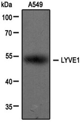
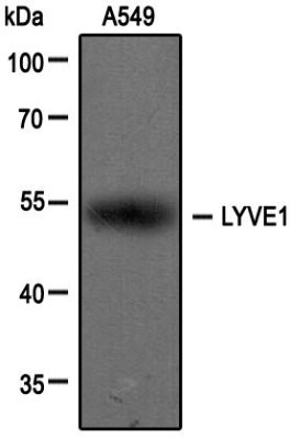
- Western Blot: LYVE-1 Antibody [NB100-725] - Analysis of extracts from A549 cells using LYVE1 antibody (NB100-725, 1:100). Image from verified customer review.
- Submitted by
- Novus Biologicals (provider)
- Main image
- Experimental details
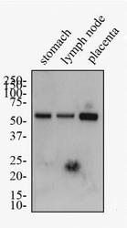
- Western Blot: LYVE-1 Antibody [NB100-725] - Western Blot: [NB100-725] - Total protein from human stomach, lymph node and placenta was separated on a 7.5% gel by SDS-PAGE, transferred to PVDF membrane and blocked in 5% non-fat milk in TBST. The membrane was probed with 1.0 ug/ml anti-LYVE1 in 1% milk, and detected with an anti-rabbit HRP secondary antibody using chemiluminescence.

Supportive validation
- Submitted by
- Novus Biologicals (provider)
- Main image
- Experimental details
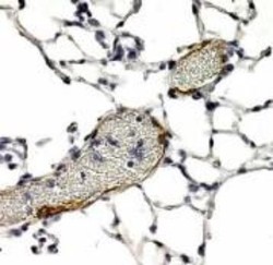
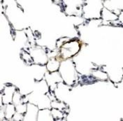
- Immunohistochemistry: LYVE-1 Antibody [NB100-725] - Detection of LYVE1 in endothelial cells of human lung blood vessels using NB100-725. Note presence of RBCs within vessel lumen.
- Submitted by
- Novus Biologicals (provider)
- Main image
- Experimental details
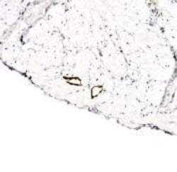
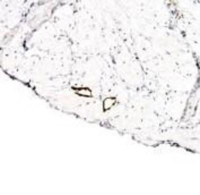
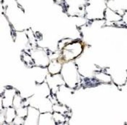
- Immunohistochemistry: LYVE-1 Antibody [NB100-725] - Detection of LYVE1 in endothelial cells of human bladder vasculature using NB100-725.
- Submitted by
- Novus Biologicals (provider)
- Main image
- Experimental details
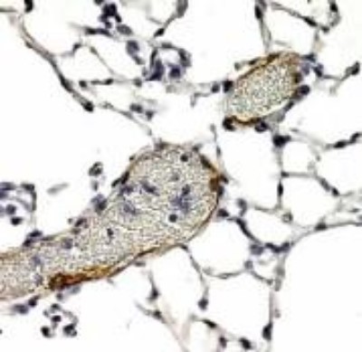
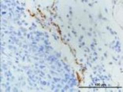
- Immunohistochemistry: LYVE-1 Antibody [NB100-725] - Detection of LYVE1 in endothelial cells of human lung blood vessels using NB100-725.
- Submitted by
- Novus Biologicals (provider)
- Main image
- Experimental details
- Immunohistochemistry-Paraffin: LYVE-1 Antibody [NB100-725] - Analysis using the DyLight 488 conjugate of NB100-725. Staining of LYVE1 in paraffin embedded MDA-MB-231 breast cancer orthotopic transplantation tissue. Image courtesy of product review submitted by Luana Schito.

Supportive validation
- Submitted by
- Novus Biologicals (provider)
- Main image
- Experimental details
- Flow Cytometry: LYVE-1 Antibody [NB100-725] - Analysis using the DyLight 488 conjugate of NB100-725. Unlabeled control with labeled samples of cells dissociated mechanically and enzymatically from human skin. Image provided via product review by Patricia Redondo.

- Submitted by
- Novus Biologicals (provider)
- Main image
- Experimental details
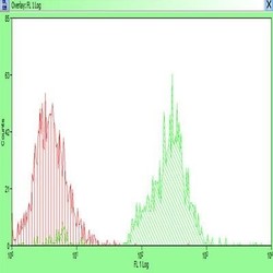
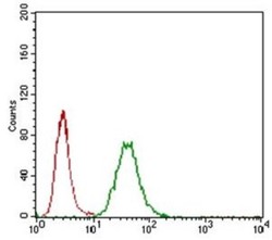
- Flow Cytometry: LYVE-1 Antibody [NB100-725] - LYVE1 antibody was tested at 1:400 in A549 cells using an Alexa Fluor 488 secondary (shown in green) alongside unstained cells (shown in red).

- Submitted by
- Novus Biologicals (provider)
- Main image
- Experimental details
- Flow Cytometry: LYVE-1 Antibody [NB100-725] - Analysis using the Alexa Fluor (R) 488 conjugate of NB100-725. Staining of LYVE1 in human dissociated skin lyve1+ using AF488 conjugated anti-LYVE1 antibody. Image from verified customer review.
- Submitted by
- Novus Biologicals (provider)
- Main image
- Experimental details
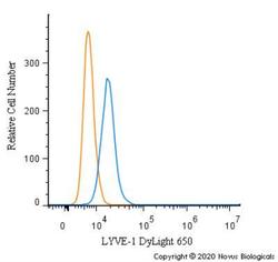
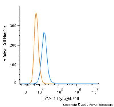
- Flow Cytometry: LYVE-1 Antibody [NB100-725] - An intracellular stain was performed on K562 cells with LYVE-1 Antibody NB100-725C (blue) and a matched isotype control (orange). Cells were fixed with 4% PFA and then permeabilized with 0.1% saponin. Cells were incubated in an antibody dilution of 2.5 ug/mL for 30 minutes at room temperature. Both antibodies were conjugated to DyLight 650.
- Submitted by
- Novus Biologicals (provider)
- Main image
- Experimental details
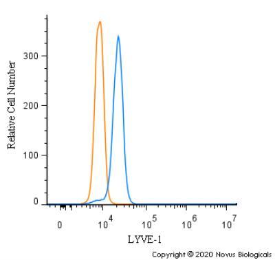
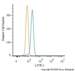
- Flow Cytometry: LYVE-1 Antibody [NB100-725] - An intracellular stain was performed on A549 cells with LYVE-1 Antibody NB100-725 (blue) and a matched isotype control (orange). Cells were fixed with 4% PFA and then permeabilized with 0.1% saponin. Cells were incubated in an antibody dilution of 1.0 ug/mL for 30 minutes at room temperature, followed by Rabbit IgG (H+L) Cross-Adsorbed Secondary Antibody, Dylight 550 (SA5-10033, Thermo Fisher).