Explore
 Validate
 Learn
LearnNBP1-89558
antibody from Novus Biologicals
Targeting: SELENOS
AD-015, MGC2553, SBBI8, SELS, SEPS1, VIMP
 Western blot
 Immunocytochemistry
Antibody data
- Antibody Data
- Antigen structure
- References [3]
- Comments [0]
- Validations
- Western blot [3]
- Immunohistochemistry [5]
Submit
Validation data
Reference
Comment
Report error
- Product number
- NBP1-89558 - Provider product page

- Provider
- Novus Biologicals
- Proper citation
- Novus Cat#NBP1-89558, RRID:AB_11024155
- Product name
- Rabbit Polyclonal SELS Antibody
- Antibody type
- Polyclonal
- Description
- Immunogen affinity purified. Specificity of human, mouse, rat SELS antibody verified on a Protein Array containing target protein plus 383 other non-specific proteins.
- Reactivity
- Human, Mouse, Rat
- Host
- Rabbit
- Isotype
- IgG
- Vial size
- 0.1 ml
- Storage
- Store at 4C short term. Aliquot and store at -20C long term. Avoid freeze-thaw cycles.
Submitted references Selective up-regulation of human selenoproteins in response to oxidative stress.
Alternative transcripts and 3'UTR elements govern the incorporation of selenocysteine into selenoprotein S.
The impact of tissue fixatives on morphology and antibody-based protein profiling in tissues and cells.
Touat-Hamici Z, Legrain Y, Bulteau AL, Chavatte L
The Journal of biological chemistry 2014 May 23;289(21):14750-61
The Journal of biological chemistry 2014 May 23;289(21):14750-61
Alternative transcripts and 3'UTR elements govern the incorporation of selenocysteine into selenoprotein S.
Bubenik JL, Miniard AC, Driscoll DM
PloS one 2013;8(4):e62102
PloS one 2013;8(4):e62102
The impact of tissue fixatives on morphology and antibody-based protein profiling in tissues and cells.
Paavilainen L, Edvinsson A, Asplund A, Hober S, Kampf C, Pontén F, Wester K
The journal of histochemistry and cytochemistry : official journal of the Histochemistry Society 2010 Mar;58(3):237-46
The journal of histochemistry and cytochemistry : official journal of the Histochemistry Society 2010 Mar;58(3):237-46
No comments: Submit comment
Supportive validation
- Submitted by
- Novus Biologicals (provider)
- Main image

- Experimental details
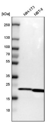
- Western Blot: SELS Antibody [NBP1-89558] - Analysis in mouse cell line NIH-3T3 and rat cell line NBT-II.
- Submitted by
- Novus Biologicals (provider)
- Main image
- Experimental details
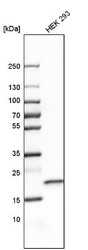
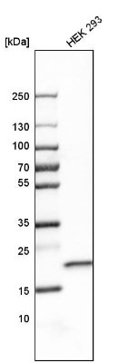
- Western Blot: SELS Antibody [NBP1-89558] - Analysis in human cell line HEK 293.
- Submitted by
- Novus Biologicals (provider)
- Main image
- Experimental details
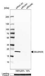
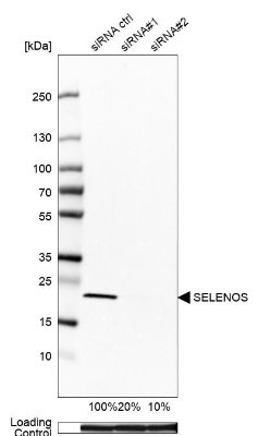
- Western Blot: SELS Antibody [NBP1-89558] - Analysis in U-251MG cells transfected with control siRNA, target specific siRNA probe #1 and #2, using Anti-SELENOS antibody. Remaining relative intensity is presented. Loading control: Anti-GAPDH.
Supportive validation
- Submitted by
- Novus Biologicals (provider)
- Main image
- Experimental details
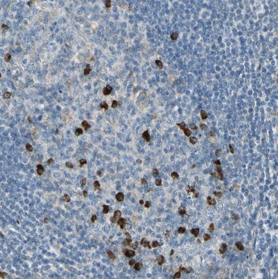
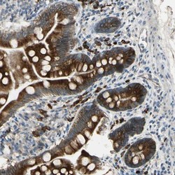
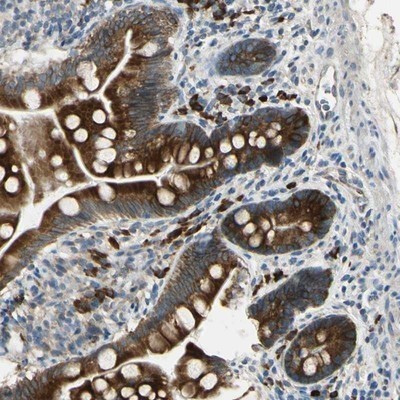
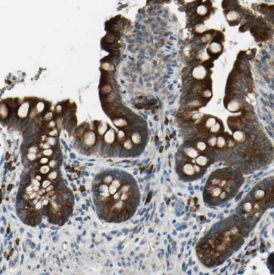
- Immunohistochemistry-Paraffin: SELS Antibody [NBP1-89558] - Staining of human small intestine shows strong cytoplasmic positivity in glandular cells.
- Submitted by
- Novus Biologicals (provider)
- Main image

- Experimental details
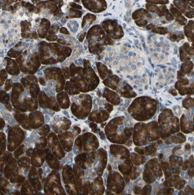
- Immunohistochemistry-Paraffin: SELS Antibody [NBP1-89558] - Staining of human pancreas shows strong cytoplasmic positivity in exocrine glandular cells.
- Submitted by
- Novus Biologicals (provider)
- Main image
- Experimental details
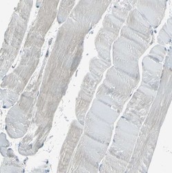
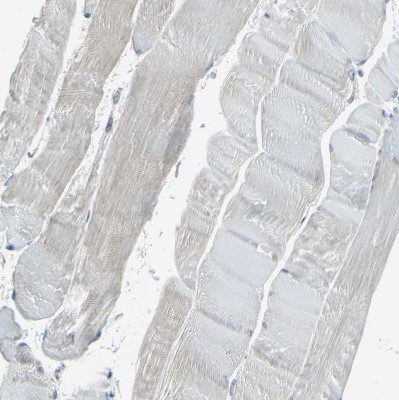
- Immunohistochemistry-Paraffin: SELS Antibody [NBP1-89558] - Staining of human skeletal muscle shows no positivity in myocytes as expected.
- Submitted by
- Novus Biologicals (provider)
- Main image

- Experimental details
- Immunohistochemistry-Paraffin: SELS Antibody [NBP1-89558] - Staining of human small intestine shows strong cytoplasmic positivity in glandular cells.
- Submitted by
- Novus Biologicals (provider)
- Main image

- Experimental details
- Immunohistochemistry-Paraffin: SELS Antibody [NBP1-89558] - Staining of human tonsil shows strong cytoplasmic positivity in a small subset of germinal center cells.