Explore
 Validate
 Learn
Learn Western blot
 Immunohistochemistry
Antibody data
- Antibody Data
- Antigen structure
- References [0]
- Comments [0]
- Validations
- Immunohistochemistry [1]
- Flow cytometry [2]
- Other assay [1]
Submit
Validation data
Reference
Comment
Report error
- Product number
- PA5-14268 - Provider product page

- Provider
- Invitrogen Antibodies
- Product name
- MOB1 Polyclonal Antibody
- Antibody type
- Polyclonal
- Antigen
- Synthetic peptide
- Description
- This antibody is predicted to react with mouse and rat based on sequence homology.
- Reactivity
- Human
- Host
- Rabbit
- Isotype
- IgG
- Vial size
- 400 μL
- Concentration
- 1.5 mg/mL
- Storage
- Store at 4°C short term. For long term storage, store at -20°C, avoiding freeze/thaw cycles.
No comments: Submit comment
Supportive validation
- Submitted by
- Invitrogen Antibodies (provider)
- Main image
- Experimental details
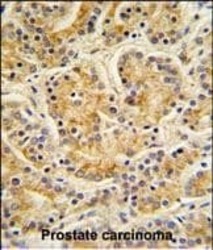
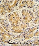
- Immunohistochemistry analysis of MOB1 in formalin fixed and paraffin embedded human prostate carcinoma. Samples were incubated with MOB1 polyclonal antibody (Product # PA5-14268) followed by peroxidase conjugation of the secondary antibody and DAB staining. This data demonstrates the use of this antibody for immunohistochemistry. Clinical relevance has not been evaluated.
Supportive validation
- Submitted by
- Invitrogen Antibodies (provider)
- Main image
- Experimental details
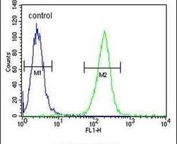
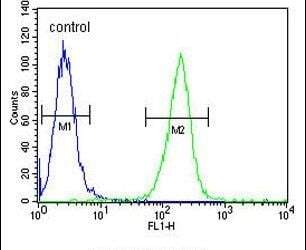
- Flow cytometry analysis of A2058 cells using a MOBKL1B polyclonal antibody (Product # PA5-14268) (right) compared to a negative control cell (left) at a dilution of 1:10-50, followed by a FITC-conjugated goat anti-rabbit antibody
- Submitted by
- Invitrogen Antibodies (provider)
- Main image
- Experimental details
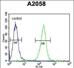
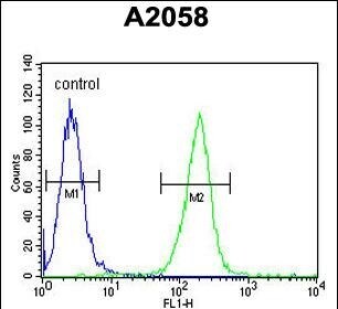
- Flow cytometry of MOB1 in A2058 cells (right histogram). Samples were incubated with MOB1 polyclonal antibody (Product # PA5-14268) followed by FITC-conjugated goat-anti-rabbit secondary antibody. Negative control cell (left histogram).
Supportive validation
- Submitted by
- Invitrogen Antibodies (provider)
- Main image

- Experimental details
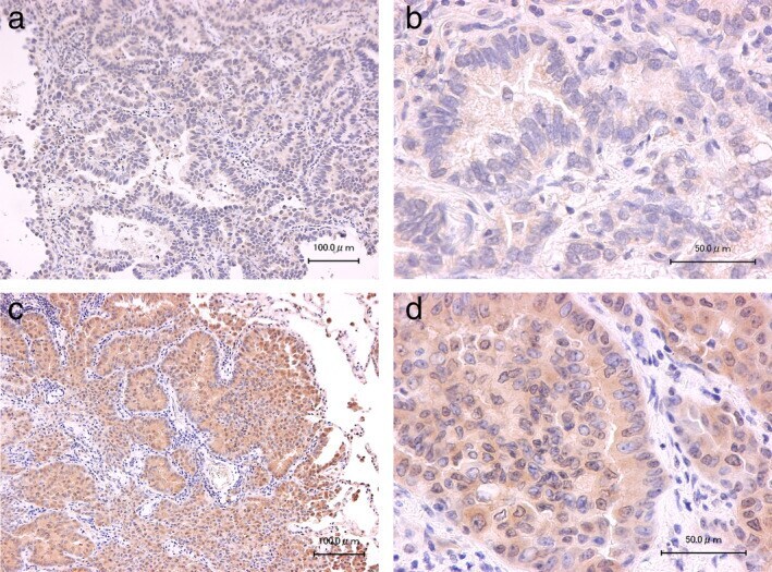
- Figure 1 Representative immunohistochemical staining of MOB1 in lung adenocarcinoma specimens. Specimens negative (score = 0) ( a and b ) or positive (score = 4) ( c and d ) for MOB1 staining are shown at low ( a and c ) and high ( b and d ) magnification. Scale bars: 100 mum ( a and c ) or 50 mum ( b and d ).