 Explore
 Validate
 Learn
Learn Western blot
 ELISA
ELISAAntibody data
- Antibody Data
- Antigen structure
- References [11]
- Comments [0]
- Validations
- Western blot [1]
- Immunocytochemistry [1]
- Immunoprecipitation [1]
Submit
Validation data
Reference
Comment
Report error
- Product number
- 14413-1-AP - Provider product page

- Provider
- Proteintech Group
- Proper citation
- Proteintech Cat#14413-1-AP, RRID:AB_10858916
- Product name
- PIBF1 antibody
- Antibody type
- Polyclonal
- Description
- KD/KO validated PIBF1 antibody (Cat. #14413-1-AP) is a rabbit polyclonal antibody that shows reactivity with human and has been validated for the following applications: IF, IP, WB,ELISA.
- Reactivity
- Human
- Host
- Rabbit
- Conjugate
- Unconjugated
- Isotype
- IgG
- Vial size
- 20ul, 150ul
Submitted references OTUD5 enhances activation of multiple cell death pathways and hyperoxia-induced lung injury by stabilizing TRAF4 and activating the p38/JNK pathway.
The Luminal Ring Protein C2CD3 Acts as a Radial In-to-Out Organizer of the Distal Centriole and Appendages.
Serotype-specific host proteome remodeling in human foreskin fibroblasts during lytic HSV-1 and HSV-2 infection.
The luminal ring protein C2CD3 acts as a radial in-to-out organizer of the distal centriole and appendages.
Multiplexed Nanoscopy via Buffer Exchange.
The evolutionary conserved proteins CEP90, FOPNL, and OFD1 recruit centriolar distal appendage proteins to initiate their assembly.
Zika virus alters centrosome organization to suppress the innate immune response.
Human SFI1 and Centrin form a complex critical for centriole architecture and ciliogenesis.
A ciliopathy complex builds distal appendages to initiate ciliogenesis.
CCDC57 Cooperates with Microtubules and Microcephaly Protein CEP63 and Regulates Centriole Duplication and Mitotic Progression.
Centriolar satellites assemble centrosomal microcephaly proteins to recruit CDK2 and promote centriole duplication.
Gu X, Zhu T, Hang G, Chen W, Chen H
Tissue & cell 2025 Oct;96:103008
Tissue & cell 2025 Oct;96:103008
The Luminal Ring Protein C2CD3 Acts as a Radial In-to-Out Organizer of the Distal Centriole and Appendages.
Bertiaux E, Louvel V, McCafferty CL, van den Hoek H, Batman U, Mukherjee S, Bournonville L, Mercey O, Mean I, Müller A, Van der Stappen P, Buss G, Daraspe J, Genoud C, Stearns T, Engel BD, Hamel V, Guichard P
bioRxiv : the preprint server for biology 2025 Jun 18;
bioRxiv : the preprint server for biology 2025 Jun 18;
Serotype-specific host proteome remodeling in human foreskin fibroblasts during lytic HSV-1 and HSV-2 infection.
Pan X, Xie J, Zhang Z, Guo X, Li J, Lin D, Qian Y, Xu J, Hu Y, Shi J
Virology journal 2025 Jul 14;22(1):239
Virology journal 2025 Jul 14;22(1):239
The luminal ring protein C2CD3 acts as a radial in-to-out organizer of the distal centriole and appendages.
Bertiaux E, Louvel V, McCafferty CL, van den Hoek H, Batman U, Mukherjee S, Bournonville L, Mercey O, Méan I, Righetto RD, Müller A, Van der Stappen P, Buss G, Daraspe J, Genoud C, Stearns T, Engel BD, Hamel V, Guichard P
PLoS biology 2025 Dec;23(12):e3003519
PLoS biology 2025 Dec;23(12):e3003519
Multiplexed Nanoscopy via Buffer Exchange.
Chang TB, Yang TT
ACS nano 2024 Aug 27;18(34):23445-23456
ACS nano 2024 Aug 27;18(34):23445-23456
The evolutionary conserved proteins CEP90, FOPNL, and OFD1 recruit centriolar distal appendage proteins to initiate their assembly.
Le Borgne P, Greibill L, Laporte MH, Lemullois M, Bouhouche K, Temagoult M, Rosnet O, Le Guennec M, Lignières L, Chevreux G, Koll F, Hamel V, Guichard P, Tassin AM
PLoS biology 2022 Sep;20(9):e3001782
PLoS biology 2022 Sep;20(9):e3001782
Zika virus alters centrosome organization to suppress the innate immune response.
Kodani A, Knopp KA, Di Lullo E, Retallack H, Kriegstein AR, DeRisi JL, Reiter JF
EMBO reports 2022 Sep 5;23(9):e52211
EMBO reports 2022 Sep 5;23(9):e52211
Human SFI1 and Centrin form a complex critical for centriole architecture and ciliogenesis.
Laporte MH, Bouhlel IB, Bertiaux E, Morrison CG, Giroud A, Borgers S, Azimzadeh J, Bornens M, Guichard P, Paoletti A, Hamel V
The EMBO journal 2022 Nov 2;41(21):e112107
The EMBO journal 2022 Nov 2;41(21):e112107
A ciliopathy complex builds distal appendages to initiate ciliogenesis.
Kumar D, Rains A, Herranz-Pérez V, Lu Q, Shi X, Swaney DL, Stevenson E, Krogan NJ, Huang B, Westlake C, Garcia-Verdugo JM, Yoder BK, Reiter JF
The Journal of cell biology 2021 Sep 6;220(9)
The Journal of cell biology 2021 Sep 6;220(9)
CCDC57 Cooperates with Microtubules and Microcephaly Protein CEP63 and Regulates Centriole Duplication and Mitotic Progression.
Gurkaslar HK, Culfa E, Arslanhan MD, Lince-Faria M, Firat-Karalar EN
Cell reports 2020 May 12;31(6):107630
Cell reports 2020 May 12;31(6):107630
Centriolar satellites assemble centrosomal microcephaly proteins to recruit CDK2 and promote centriole duplication.
Kodani A, Yu TW, Johnson JR, Jayaraman D, Johnson TL, Al-Gazali L, Sztriha L, Partlow JN, Kim H, Krup AL, Dammermann A, Krogan NJ, Walsh CA, Reiter JF
eLife 2015 Aug 22;4
eLife 2015 Aug 22;4
No comments: Submit comment
Supportive validation
- Submitted by
- Proteintech Group (provider)
- Main image

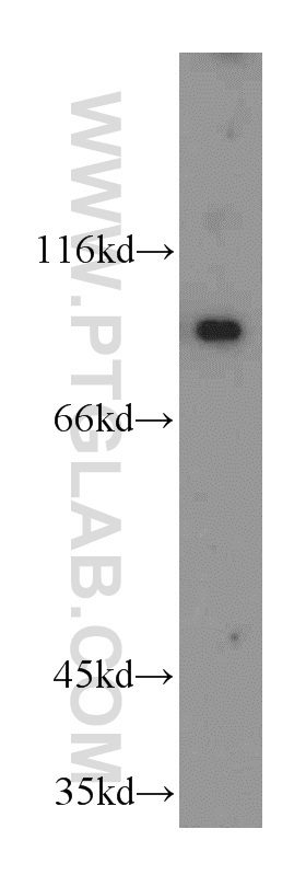
- Experimental details
- HEK-293 cells were subjected to SDS PAGE followed by western blot with 14413-1-AP(PIBF1 antibody) at dilution of 1:500
- Sample type
- cell line
Supportive validation
- Submitted by
- Proteintech Group (provider)
- Main image
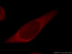
- Experimental details
- Immunofluorescent analysis of MCF-7 cells, using PIBF1 antibody 14413-1-AP at 1:25 dilution and Rhodamine-labeled goat anti-rabbit IgG (red).
- Sample type
- cell line

Supportive validation
- Submitted by
- Proteintech Group (provider)
- Main image
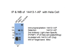
- Experimental details
- IP result of anti-PIBF1(14413-1-AP for IP and Detection).
- Sample type
- cell line
