Explore
 Validate
 Learn
LearnPA5-21429
antibody from Invitrogen Antibodies
Targeting: ARHGEF18
KIAA0521, MGC15913, P114-RhoGEF, p114RhoGEF
 Western blot
 Immunocytochemistry
Antibody data
- Antibody Data
- Antigen structure
- References [1]
- Comments [0]
- Validations
- Immunocytochemistry [4]
- Immunohistochemistry [2]
- Other assay [1]
Submit
Validation data
Reference
Comment
Report error
- Product number
- PA5-21429 - Provider product page

- Provider
- Invitrogen Antibodies
- Product name
- ARHGEF18 Polyclonal Antibody
- Antibody type
- Polyclonal
- Antigen
- Recombinant full-length protein
- Description
- Recommended positive controls: HepG2. Predicted reactivity: Mouse (89%), Rat (89%). Store product as a concentrated solution. Centrifuge briefly prior to opening the vial.
- Reactivity
- Human, Mouse, Rat
- Host
- Rabbit
- Isotype
- IgG
- Vial size
- 100 μL
- Concentration
- 1.0 mg/mL
- Storage
- Store at 4°C short term. For long term storage, store at -20°C, avoiding freeze/thaw cycles.
Submitted references GPCR-independent activation of G proteins promotes apical cell constriction in vivo.
Marivin A, Morozova V, Walawalkar I, Leyme A, Kretov DA, Cifuentes D, Dominguez I, Garcia-Marcos M
The Journal of cell biology 2019 May 6;218(5):1743-1763
The Journal of cell biology 2019 May 6;218(5):1743-1763
No comments: Submit comment
Supportive validation
- Submitted by
- Invitrogen Antibodies (provider)
- Main image

- Experimental details
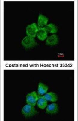
- Immunofluorescent analysis of ARHGEF18 in methanol-fixed A431 cells using an ARHGEF18 polyclonal antibody (Product # PA5-21429) at a 1:200 dilution.
- Submitted by
- Invitrogen Antibodies (provider)
- Main image

- Experimental details
- Immunofluorescence analysis of methanol-fixed A431, using ARHGEF18 antibody (Product # PA5-21429) at 1:200 dilution.

- Submitted by
- Invitrogen Antibodies (provider)
- Main image
- Experimental details
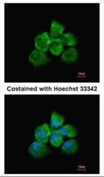
- Immunofluorescence analysis of methanol-fixed A431, using ARHGEF18 antibody (Product # PA5-21429) at 1:200 dilution.

- Submitted by
- Invitrogen Antibodies (provider)
- Main image

- Experimental details
- Immunofluorescence analysis of methanol-fixed A431, using ARHGEF18 antibody (Product # PA5-21429) at 1:200 dilution.

Supportive validation
- Submitted by
- Invitrogen Antibodies (provider)
- Main image

- Experimental details
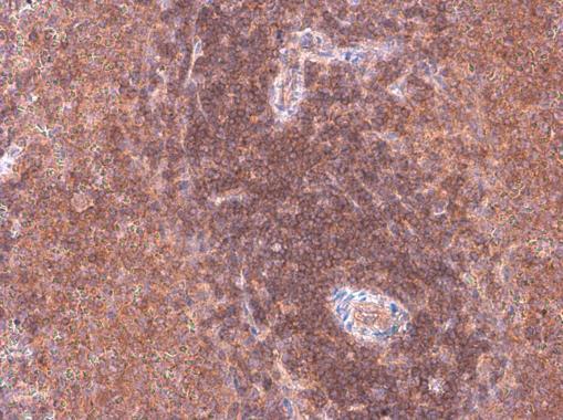
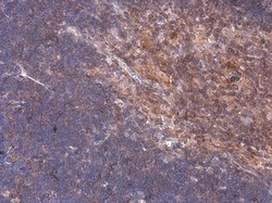
- Immunohistochemistry (Paraffin) analysis of ARHGEF18 was performed in paraffin-embedded rat spleen tissue using ARHGEF18 Polyclonal Antibody (Product # PA5-21429) at a dilution of 1:500.
- Submitted by
- Invitrogen Antibodies (provider)
- Main image
- Experimental details
- Immunohistochemistry (Paraffin) analysis of ARHGEF18 was performed in paraffin-embedded mouse thymus gland tissue using ARHGEF18 Polyclonal Antibody (Product # PA5-21429) at a dilution of 1:500.

Supportive validation
- Submitted by
- Invitrogen Antibodies (provider)
- Main image
- Experimental details
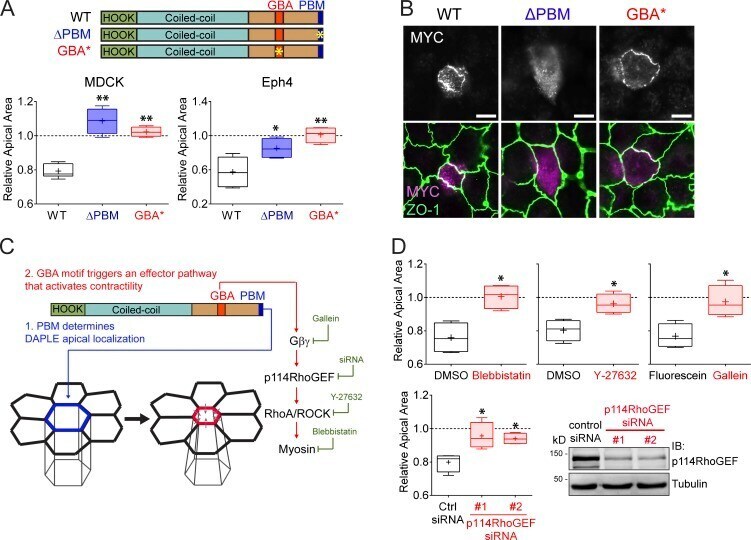
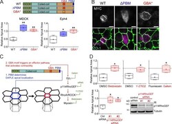
- Both the PBM and GBA motif of DAPLE are required to promote apical cell constriction. (A) Quantification of the relative apical area of DAPLE-transfected cells compared with neighboring, untransfected cells shows that hDAPLE ΔPBM and hDAPLE GBA* mutants fail to promote apical constriction in MDCK (left) or EpH4 (right) cells. Results are presented as box-and-whiskers plots (error bars indicate minimum to maximum range) of n = 4-9 independent experiments per condition. *, P < 0.05; **, P < 0.01 using the Mann-Whitney U test. (B) Confocal fluorescence microscopy pictures of MDCK cells sparsely expressing the indicated MYC-hDAPLE constructs and costained for MYC (magenta) and ZO-1 (green) show that hDAPLE GBA* mutant is enriched at apical junctions like WT, while hDAPLE ΔPBM mutant is not. Scale bars, 10 µm. (C) Diagram depicting the different functions of the PBM and the GBA motif in DAPLE-induced apical cell constriction. A possible effector pathway to promote apical cell constriction though G protein activation is shown on the right, along with the treatments used in D to test it (green). (D) Box-and-whiskers plots (error bars indicate minimum to maximum range) for the quantification of relative apical area show that DAPLE-mediated apical constriction requires the activity of myosin (inhibited by blebbistatin), ROCK (inhibited by Y-27632), free Gßγ (inhibited by Gallein, but not its inactive analogue, fluorescein) and p114RhoGEF (inhibited by siRNA). n = 4 independent experim