Explore
 Validate
 Learn
Learn Western blot
Antibody data
- Antibody Data
- Antigen structure
- References [1]
- Comments [0]
- Validations
- Western blot [1]
- Immunocytochemistry [7]
- Immunohistochemistry [7]
- Flow cytometry [2]
- Other assay [1]
Submit
Validation data
Reference
Comment
Report error
- Product number
- MA5-34952 - Provider product page

- Provider
- Invitrogen Antibodies
- Product name
- SPATA5L1 Monoclonal Antibody (16A2)
- Antibody type
- Monoclonal
- Antigen
- Recombinant full-length protein
- Description
- Positive Control: K562 cell lysates, HCT116, JAR, MG-63, rat seminal vesicle tissue, human skin tissue, human breast carcinoma tissue, human placenta tissue, human small intestine tissue.
- Reactivity
- Human
- Host
- Mouse
- Isotype
- IgG
- Antibody clone number
- 16A2
- Vial size
- 100 μL
- Concentration
- 2 mg/mL
- Storage
- -20°C, Avoid Freeze/Thaw Cycles, store in dark
Submitted references Labeling of heterochronic ribosomes reveals C1ORF109 and SPATA5 control a late step in human ribosome assembly.
Ni C, Schmitz DA, Lee J, Pawłowski K, Wu J, Buszczak M
Cell reports 2022 Mar 29;38(13):110597
Cell reports 2022 Mar 29;38(13):110597
No comments: Submit comment
Supportive validation
- Submitted by
- Invitrogen Antibodies (provider)
- Main image

- Experimental details
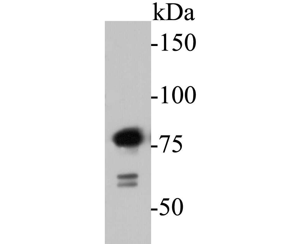
- Western blot analysis of SPATA5L1 in K562 cell lysate. Samples were transferred to PVDF membrane, blocked with 5% BSA (1 hour), incubated with SPATA5L1 monoclonal antibody (Product # MA5-34952), at a dilution of 1:500, followed by Goat Anti-Rabbit IgG-HRP (1 hour) with a dilution of 1:5000.
Supportive validation
- Submitted by
- Invitrogen Antibodies (provider)
- Main image

- Experimental details
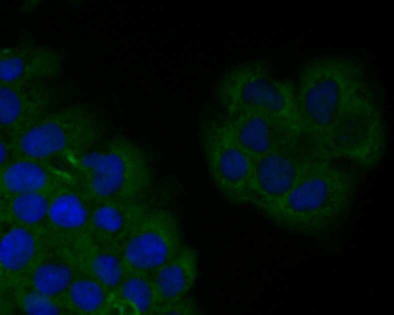
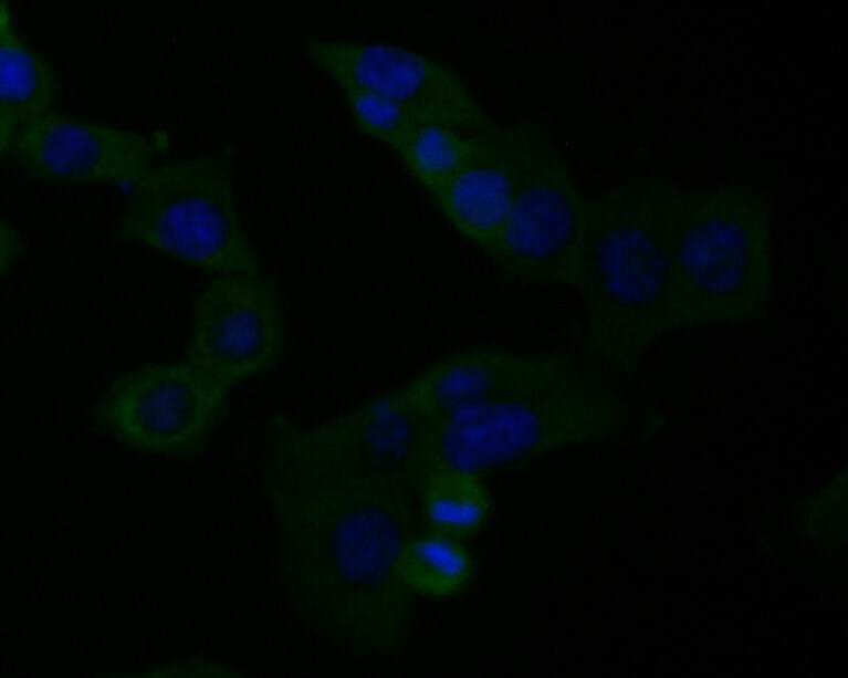
- Immunofluorescent analysis of SPATA5L1 in JAR cells (green). Samples were formalin fixed, permeabilized with 0.1% Triton X-100 in TBS (1 hour, room temperature) and blocked with 1% BSA (15 min, room temperature), incubated with SPATA5L1 monoclonal antibody (Product # MA5-34952) at a dilution of 1:50 (1 hour, room temperature), and followed by Alexa Fluor 488 Goat anti-Rabbit IgG and DAPI (blue) with a dilution of 1:1000.
- Submitted by
- Invitrogen Antibodies (provider)
- Main image

- Experimental details
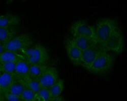
- Immunofluorescent analysis of SPATA5L1 in MG-63 cells (green). Samples were formalin fixed, permeabilized with 0.1% Triton X-100 in TBS (1 hour, room temperature) and blocked with 1% BSA (15 min, room temperature), incubated with SPATA5L1 monoclonal antibody (Product # MA5-34952) at a dilution of 1:50 (1 hour, room temperature), and followed by Alexa Fluor 488 Goat anti-Rabbit IgG and DAPI (blue) with a dilution of 1:1000.

- Submitted by
- Invitrogen Antibodies (provider)
- Main image

- Experimental details
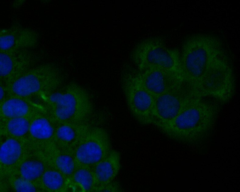
- Immunofluorescent analysis of SPATA5L1 in JAR cells (green). Samples were formalin fixed, permeabilized with 0.1% Triton X-100 in TBS (1 hour, room temperature) and blocked with 1% BSA (15 min, room temperature), incubated with SPATA5L1 monoclonal antibody (Product # MA5-34952) at a dilution of 1:50 (1 hour, room temperature), and followed by Alexa Fluor 488 Goat anti-Rabbit IgG and DAPI (blue) with a dilution of 1:1000.

- Submitted by
- Invitrogen Antibodies (provider)
- Main image
- Experimental details
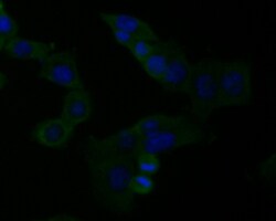
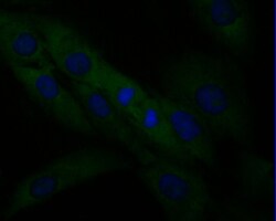
- Immunofluorescent analysis of SPATA5L1 in HCT116 cells (green). Samples were formalin fixed, permeabilized with 0.1% Triton X-100 in TBS (1 hour, room temperature) and blocked with 1% BSA (15 min, room temperature), incubated with SPATA5L1 monoclonal antibody (Product # MA5-34952) at a dilution of 1:50 (1 hour, room temperature), and followed by Alexa Fluor 488 Goat anti-Rabbit IgG and DAPI (blue) with a dilution of 1:1000.

- Submitted by
- Invitrogen Antibodies (provider)
- Main image

- Experimental details
- Immunofluorescent analysis of SPATA5L1 in HCT116 cells (green). Samples were formalin fixed, permeabilized with 0.1% Triton X-100 in TBS (1 hour, room temperature) and blocked with 1% BSA (15 min, room temperature), incubated with SPATA5L1 monoclonal antibody (Product # MA5-34952) at a dilution of 1:50 (1 hour, room temperature), and followed by Alexa Fluor 488 Goat anti-Rabbit IgG and DAPI (blue) with a dilution of 1:1000.
- Submitted by
- Invitrogen Antibodies (provider)
- Main image
- Experimental details
- Immunofluorescent analysis of SPATA5L1 in JAR cells (green). Samples were formalin fixed, permeabilized with 0.1% Triton X-100 in TBS (1 hour, room temperature) and blocked with 1% BSA (15 min, room temperature), incubated with SPATA5L1 monoclonal antibody (Product # MA5-34952) at a dilution of 1:50 (1 hour, room temperature), and followed by Alexa Fluor 488 Goat anti-Rabbit IgG and DAPI (blue) with a dilution of 1:1000.
- Submitted by
- Invitrogen Antibodies (provider)
- Main image
- Experimental details
- Immunofluorescent analysis of SPATA5L1 in MG-63 cells (green). Samples were formalin fixed, permeabilized with 0.1% Triton X-100 in TBS (1 hour, room temperature) and blocked with 1% BSA (15 min, room temperature), incubated with SPATA5L1 monoclonal antibody (Product # MA5-34952) at a dilution of 1:50 (1 hour, room temperature), and followed by Alexa Fluor 488 Goat anti-Rabbit IgG and DAPI (blue) with a dilution of 1:1000.

Supportive validation
- Submitted by
- Invitrogen Antibodies (provider)
- Main image

- Experimental details
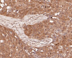
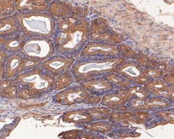
- Immunohistochemistry analysis of SPATA5L1 in paraffin-embedded rat seminal vesicle tissue. Samples were heat mediated antigen retrieval with Tris-EDTA buffer (pH 8.0-8.4, 20 minutes) and blocked in 5% BSA (30 min, room temperature), incubated with SPATA5L1 monoclonal antibody (Product # MA5-34952) at a dilution of 1:50 (30 min, room temperature), and followed by HRP conjugate, DAB and hematoxylin (mounted with DPX).

- Submitted by
- Invitrogen Antibodies (provider)
- Main image
- Experimental details
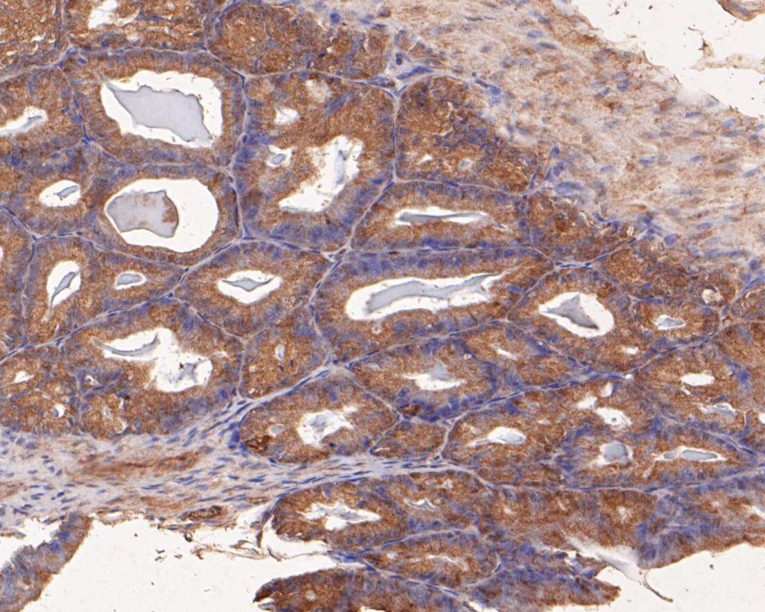
- Immunohistochemistry analysis of SPATA5L1 in paraffin-embedded human breast carcinoma tissue. Samples were heat mediated antigen retrieval with Tris-EDTA buffer (pH 8.0-8.4, 20 minutes) and blocked in 5% BSA (30 min, room temperature), incubated with SPATA5L1 monoclonal antibody (Product # MA5-34952) at a dilution of 1:50 (30 min, room temperature), and followed by HRP conjugate, DAB and hematoxylin (mounted with DPX).

- Submitted by
- Invitrogen Antibodies (provider)
- Main image
- Experimental details
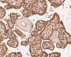
- Immunohistochemistry analysis of SPATA5L1 in paraffin-embedded human placenta tissue. Samples were heat mediated antigen retrieval with Tris-EDTA buffer (pH 8.0-8.4, 20 minutes) and blocked in 5% BSA (30 min, room temperature), incubated with SPATA5L1 monoclonal antibody (Product # MA5-34952) at a dilution of 1:50 (30 min, room temperature), and followed by HRP conjugate, DAB and hematoxylin (mounted with DPX).

- Submitted by
- Invitrogen Antibodies (provider)
- Main image

- Experimental details
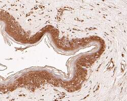
- Immunohistochemistry analysis of SPATA5L1 in paraffin-embedded human skin tissue. Samples were heat mediated antigen retrieval with Tris-EDTA buffer (pH 8.0-8.4, 20 minutes) and blocked in 5% BSA (30 min, room temperature), incubated with SPATA5L1 monoclonal antibody (Product # MA5-34952) at a dilution of 1:50 (30 min, room temperature), and followed by HRP conjugate, DAB and hematoxylin (mounted with DPX).

- Submitted by
- Invitrogen Antibodies (provider)
- Main image
- Experimental details
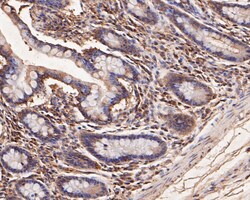
- Immunohistochemistry analysis of SPATA5L1 in paraffin-embedded human small intestine tissue. Samples were heat mediated antigen retrieval with Tris-EDTA buffer (pH 8.0-8.4, 20 minutes) and blocked in 5% BSA (30 min, room temperature), incubated with SPATA5L1 monoclonal antibody (Product # MA5-34952) at a dilution of 1:50 (30 min, room temperature), and followed by HRP conjugate, DAB and hematoxylin (mounted with DPX).

- Submitted by
- Invitrogen Antibodies (provider)
- Main image
- Experimental details
- Immunohistochemistry analysis of SPATA5L1 in paraffin-embedded human skin tissue. Samples were heat mediated antigen retrieval with Tris-EDTA buffer (pH 8.0-8.4, 20 minutes) and blocked in 5% BSA (30 min, room temperature), incubated with SPATA5L1 monoclonal antibody (Product # MA5-34952) at a dilution of 1:50 (30 min, room temperature), and followed by HRP conjugate, DAB and hematoxylin (mounted with DPX).

- Submitted by
- Invitrogen Antibodies (provider)
- Main image
- Experimental details
- Immunohistochemistry analysis of SPATA5L1 in paraffin-embedded rat seminal vesicle tissue. Samples were heat mediated antigen retrieval with Tris-EDTA buffer (pH 8.0-8.4, 20 minutes) and blocked in 5% BSA (30 min, room temperature), incubated with SPATA5L1 monoclonal antibody (Product # MA5-34952) at a dilution of 1:50 (30 min, room temperature), and followed by HRP conjugate, DAB and hematoxylin (mounted with DPX).
Supportive validation
- Submitted by
- Invitrogen Antibodies (provider)
- Main image

- Experimental details
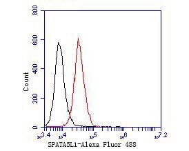
- Flow cytometry of SPATA5L1 in JAR cells, unlabelled sample (control; cells without incubation with primary antibody; black). Samples were incubated with SPATA5L1 monoclonal antibody (Product # MA5-34952) at a dilution of 1:50, followed by Alexa Fluor 488-conjugated Goat anti-Mouse IgG with a dilution of 1:1000 (30 min).
- Submitted by
- Invitrogen Antibodies (provider)
- Main image
- Experimental details
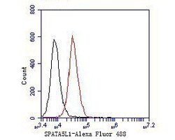
- Flow cytometry of SPATA5L1 in JAR cells, unlabelled sample (control; cells without incubation with primary antibody; black). Samples were incubated with SPATA5L1 monoclonal antibody (Product # MA5-34952) at a dilution of 1:50, followed by Alexa Fluor 488-conjugated Goat anti-Mouse IgG with a dilution of 1:1000 (30 min).

Supportive validation
- Submitted by
- Invitrogen Antibodies (provider)
- Main image
- Experimental details
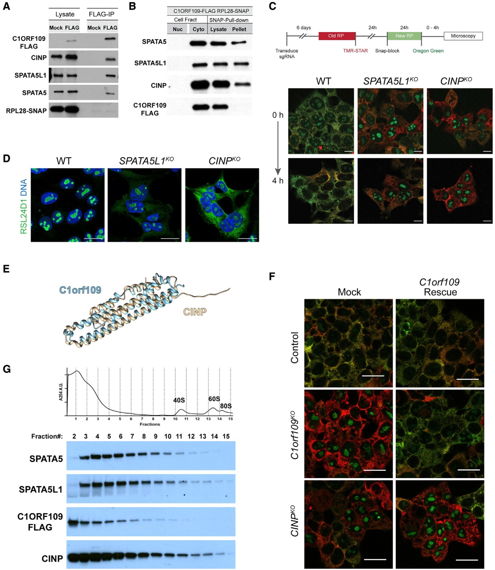
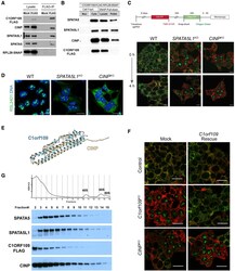
- Figure 6. SPATA5 and C1ORF109 interact with CINP and SPATA5L1 (A) Immunoprecipitation of FLAG-tagged C1ORF109 from HEK293T cell extracts crosslinked with DSP reveals that C1ORF109 physically interacts with SPATA5, SPATA5L1, and CINP, but not with RPL28. (B) Pull-down of SNAP-tagged RPL28 shows that SPATA5, SPATA5L1, and CINP interact with ribosomes, but C1ORF109 does not. (C) Knockout of SPATA5L1 or CINP leads to defects in the trafficking of newly labeled RPL28 from the nucleolus to the cytoplasm. (D) Control, SPATA5L1 KO , and C1orf109 KO cells stained for RSL24D1 (green) and DNA (blue). (E) Matchmaker alignment of CINP and C1ORF109 AlphaFold predicted structures. (F) Control, CINP KO , and C1orf109 KO cells transfected with a control or C1orf109 rescue construct stained for old and new ribosomes. (G) Cytoplasmic lysates from C1ORF109-3xFLAG expressing cells subjected to sucrose gradient fractionation and probed for SPATA5, SPATA5L1, C1ORF109, and CINP.