 Explore
 Validate
 Learn
Learn Western blot
 Immunocytochemistry
 Immunoprecipitation
Antibody data
- Antibody Data
- Antigen structure
- References [1]
- Comments [0]
- Validations
- Immunocytochemistry [5]
- Immunohistochemistry [3]
Submit
Validation data
Reference
Comment
Report error
- Product number
- PA5-27876 - Provider product page

- Provider
- Invitrogen Antibodies
- Product name
- KLF5 Polyclonal Antibody
- Antibody type
- Polyclonal
- Antigen
- Recombinant full-length protein
- Description
- Recommended positive controls: A549, HCT116, KLF5-transfected 293T. Predicted reactivity: Mouse (89%), Rat (90%), Pig (96%), Bovine (96%). Store product as a concentrated solution. Centrifuge briefly prior to opening the vial.
- Reactivity
- Human, Mouse, Rat
- Host
- Rabbit
- Isotype
- IgG
- Vial size
- 100 μL
- Concentration
- 0.5 mg/mL
- Storage
- Store at 4°C short term. For long term storage, store at -20°C, avoiding freeze/thaw cycles.
Submitted references Epigenetic reader BRD4 supports mycobacterial pathogenesis by co-modulating host lipophagy and angiogenesis.
Mukherjee T, Bhatt B, Prakhar P, Lohia GK, Rajmani RS, Balaji KN
Autophagy 2022 Feb;18(2):391-408
Autophagy 2022 Feb;18(2):391-408
No comments: Submit comment
Supportive validation
- Submitted by
- Invitrogen Antibodies (provider)
- Main image
- Experimental details
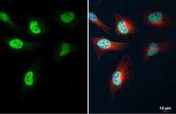
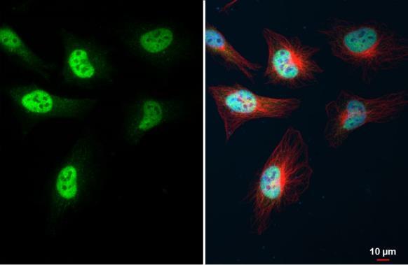
- KLF5 Polyclonal Antibody detects KLF5 protein at nucleus by immunofluorescent analysis. Sample: HeLa cells were fixed in 4% paraformaldehyde at RT for 15 min. Green: KLF5 stained by KLF5 Polyclonal Antibody (Product # PA5-27876) diluted at 1:500. Red: alpha Tubulin, a cytoskeleton marker, stained by alpha Tubulin Polyclonal Antibody [GT114] (Product # MA5-31466) diluted at 1:1,000. Blue: Fluoroshield with DAPI .
- Submitted by
- Invitrogen Antibodies (provider)
- Main image
- Experimental details
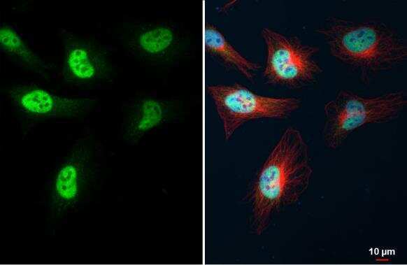
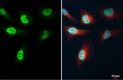
- Immunocytochemistry-Immunofluorescence analysis of KLF5 was performed in HeLa cells fixed in 4% paraformaldehyde at RT for 15 min. Green: KLF5 Polyclonal Antibody (Product # PA5-27876) diluted at 1:500. Red: Phalloidin, a cytoskeleton marker. Scale bar = 10 µm.
- Submitted by
- Invitrogen Antibodies (provider)
- Main image

- Experimental details
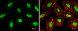
- KLF5 Polyclonal Antibody detects KLF5 protein at nucleus by immunofluorescent analysis. Sample: HeLa cells were fixed in 4% paraformaldehyde at RT for 15 min. Green: KLF5 stained by KLF5 Polyclonal Antibody (Product # PA5-27876) diluted at 1:500. Red: alpha Tubulin, a cytoskeleton marker, stained by alpha Tubulin Polyclonal Antibody [GT114] (Product # MA5-31466) diluted at 1:1,000. Blue: Fluoroshield with DAPI .
- Submitted by
- Invitrogen Antibodies (provider)
- Main image
- Experimental details
- KLF5 Polyclonal Antibody detects KLF5 protein at nucleus by immunofluorescent analysis. Sample: HeLa cells were fixed in 4% paraformaldehyde at RT for 15 min. Green: KLF5 stained by KLF5 Polyclonal Antibody (Product # PA5-27876) diluted at 1:500. Red: alpha Tubulin, a cytoskeleton marker, stained by alpha Tubulin Polyclonal Antibody [GT114] (Product # MA5-31466) diluted at 1:1,000. Blue: Fluoroshield with DAPI .

- Submitted by
- Invitrogen Antibodies (provider)
- Main image

- Experimental details
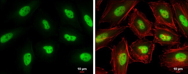
- Immunocytochemistry-Immunofluorescence analysis of KLF5 was performed in HeLa cells fixed in 4% paraformaldehyde at RT for 15 min. Green: KLF5 Polyclonal Antibody (Product # PA5-27876) diluted at 1:500. Red: Phalloidin, a cytoskeleton marker. Scale bar = 10 µm.

Supportive validation
- Submitted by
- Invitrogen Antibodies (provider)
- Main image
- Experimental details
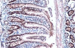
- KLF5 Polyclonal Antibody detects KLF5 protein at nucleus by immunohistochemical analysis. Sample: Paraffin-embedded mouse intestine. KLF5 stained by KLF5 Polyclonal Antibody (Product # PA5-27876) diluted at 1:500. Antigen Retrieval: Citrate buffer, pH 6.0, 15 min.

- Submitted by
- Invitrogen Antibodies (provider)
- Main image
- Experimental details
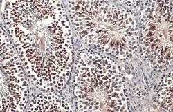
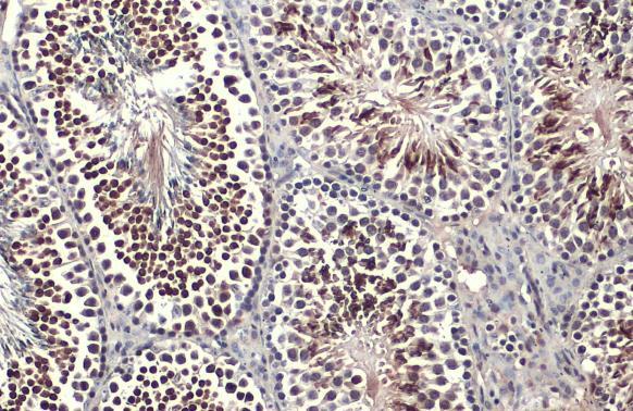
- KLF5 Polyclonal Antibody detects KLF5 protein at nucleus by immunohistochemical analysis. Sample: Paraffin-embedded mouse testis. KLF5 stained by KLF5 Polyclonal Antibody (Product # PA5-27876) diluted at 1:500. Antigen Retrieval: Citrate buffer, pH 6.0, 15 min.
- Submitted by
- Invitrogen Antibodies (provider)
- Main image
- Experimental details
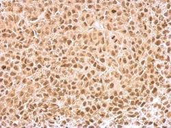
- KLF5 Polyclonal Antibody detects KLF5 protein at nucleus on HeLa xenograft by immunohistochemical analysis. Sample: Paraffin-embedded HeLa xenograft. KLF5 Polyclonal Antibody (Product # PA5-27876) dilution: 1:500. Antigen Retrieval: EDTA based buffer, pH 8.0, 15 min.
