 Explore
 Validate
 Learn
Learn Western blot
Antibody data
- Antibody Data
- Antigen structure
- References [4]
- Comments [0]
- Validations
- Western blot [3]
- Immunocytochemistry [1]
Submit
Validation data
Reference
Comment
Report error
- Product number
- AF3758 - Provider product page

- Provider
- R&D Systems
- Product name
- Human/Mouse KLF5 Antibody
- Antibody type
- Polyclonal
- Description
- Antigen Affinity-purified. Detects human and mouse KLF5 in Western blots. In direct ELISAs and Western blots, less than 5% cross-reactivity with recombinant human (rh) KLF4, rhKLF6, recombinant mouse (rm) KLF4 and rmKLF15 is observed.
- Reactivity
- Human, Mouse
- Host
- Goat
- Conjugate
- Unconjugated
- Antigen sequence
Q13887- Isotype
- IgG
- Vial size
- 100 ug
- Concentration
- LYOPH
- Storage
- Use a manual defrost freezer and avoid repeated freeze-thaw cycles. 12 months from date of receipt, -20 to -70 °C as supplied. 1 month, 2 to 8 °C under sterile conditions after reconstitution. 6 months, -20 to -70 °C under sterile conditions after reconstitution.
Submitted references Stem Cell Lineage Infidelity Drives Wound Repair and Cancer.
KLF13 regulates the differentiation-dependent human papillomavirus life cycle in keratinocytes through STAT5 and IL-8.
The intestinal epithelial cell differentiation marker intestinal alkaline phosphatase (ALPi) is selectively induced by histone deacetylase inhibitors (HDACi) in colon cancer cells in a Kruppel-like factor 5 (KLF5)-dependent manner.
Epithelial microRNAs regulate gut mucosal immunity via epithelium-T cell crosstalk.
Ge Y, Gomez NC, Adam RC, Nikolova M, Yang H, Verma A, Lu CP, Polak L, Yuan S, Elemento O, Fuchs E
Cell 2017 May 4;169(4):636-650.e14
Cell 2017 May 4;169(4):636-650.e14
KLF13 regulates the differentiation-dependent human papillomavirus life cycle in keratinocytes through STAT5 and IL-8.
Zhang W, Hong S, Maniar KP, Cheng S, Jie C, Rademaker AW, Krensky AM, Clayberger C
Oncogene 2016 Oct 20;35(42):5565-5575
Oncogene 2016 Oct 20;35(42):5565-5575
The intestinal epithelial cell differentiation marker intestinal alkaline phosphatase (ALPi) is selectively induced by histone deacetylase inhibitors (HDACi) in colon cancer cells in a Kruppel-like factor 5 (KLF5)-dependent manner.
Shin J, Carr A, Corner GA, Tögel L, Dávalos-Salas M, Tran H, Chueh AC, Al-Obaidi S, Chionh F, Ahmed N, Buchanan DD, Young JP, Malo MS, Hodin RA, Arango D, Sieber OM, Augenlicht LH, Dhillon AS, Weber TK, Mariadason JM
The Journal of biological chemistry 2014 Sep 5;289(36):25306-16
The Journal of biological chemistry 2014 Sep 5;289(36):25306-16
Epithelial microRNAs regulate gut mucosal immunity via epithelium-T cell crosstalk.
Biton M, Levin A, Slyper M, Alkalay I, Horwitz E, Mor H, Kredo-Russo S, Avnit-Sagi T, Cojocaru G, Zreik F, Bentwich Z, Poy MN, Artis D, Walker MD, Hornstein E, Pikarsky E, Ben-Neriah Y
Nature immunology 2011 Mar;12(3):239-46
Nature immunology 2011 Mar;12(3):239-46
No comments: Submit comment
Supportive validation
- Submitted by
- R&D Systems (provider)
- Main image
- Experimental details
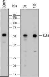
- Detection of Human and Mouse KLF5 by Western Blot. Western blot shows lysates of BG01V human embryonic stem cells, D3 mouse embryonic stem cell line, and P19 mouse embryonal carcinoma cell line. PVDF membrane was probed with 1 µg/mL of Goat Anti-Human KLF5 Antigen Affinity-purified Polyclonal Antibody (Catalog # AF3758) followed by HRP-conjugated Anti-Goat IgG Secondary Antibody (Catalog # HAF109). A specific band was detected for KLF5 at approximately 50-55 kDa (as indicated). This experiment was conducted under reducing conditions and using Immunoblot Buffer Group 1.

- Submitted by
- R&D Systems (provider)
- Main image
- Experimental details
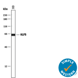
- Detection of Mouse KLF5 by Simple WesternTM. Simple Western lane view shows lysates of D3 mouse embryonic stem cell line, loaded at 0.2 mg/mL. A specific band was detected for KLF5 at approximately 64 kDa (as indicated) using 10 µg/mL of Goat Anti-Human/Mouse KLF5 Antigen Affinity-purified Polyclonal Antibody (Catalog # AF3758) followed by 1:50 dilution of HRP-conjugated Anti-Goat IgG Secondary Antibody (Catalog # HAF109). This experiment was conducted under reducing conditions and using the 12-230 kDa separation system.

- Submitted by
- R&D Systems (provider)
- Main image
- Experimental details
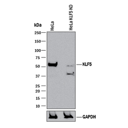
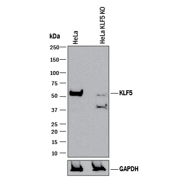
- Western Blot Shows Human KLF5 Specificity by Using Knockout Cell Line. Western blot shows lysates of HeLa human cervical epithelial carcinoma parental cell line and KLF5 knockout HeLa cell line (KO). PVDF membrane was probed with 1 µg/mL of Goat Anti-Human/Mouse KLF5 Antigen Affinity-purified Polyclonal Antibody (Catalog # AF3758) followed by HRP-conjugated Anti-Goat IgG Secondary Antibody (Catalog # HAF017). A specific band was detected for KLF5 at approximately 53 kDa (as indicated) in the parental HeLa cell line, but is not detectable in knockout HeLa cell line. GAPDH (Catalog # AF5718) is shown as a loading control. This experiment was conducted under reducing conditions and using Immunoblot Buffer Group 1.
Supportive validation
- Submitted by
- R&D Systems (provider)
- Main image
- Experimental details
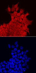
- KLF5 in D3 Mouse Stem Cells. KLF5 was detected in immersion fixed D3 mouse embryonic stem cell line using Goat Anti-Human KLF5 Antigen Affinity-purified Polyclonal Antibody (Catalog # AF3758) at 10 µg/mL for 3 hours at room temperature. Cells were stained using the NorthernLights™ 557-conjugated Anti-Goat IgG Secondary Antibody (red, upper panel; Catalog # NL001) and counterstained with DAPI (blue, lower panel). Specific staining was localized to nuclei and cytoplasm. View our protocol for Fluorescent ICC Staining of Cells on Coverslips.
