Explore
 Validate
 Learn
Learn Western blot
 ELISA
ELISAAntibody data
- Antibody Data
- Antigen structure
- References [6]
- Comments [0]
- Validations
- Western blot [1]
- Immunocytochemistry [1]
- Immunohistochemistry [2]
Submit
Validation data
Reference
Comment
Report error
- Product number
- 14833-1-AP - Provider product page

- Provider
- Proteintech Group
- Proper citation
- Proteintech Cat#14833-1-AP, RRID:AB_10859775
- Product name
- GLYR1 antibody
- Antibody type
- Polyclonal
- Description
- KD/KO validated GLYR1 antibody (Cat. #14833-1-AP) is a rabbit polyclonal antibody that shows reactivity with human, mouse, rat and has been validated for the following applications: IF, IHC, IP, WB,ELISA.
- Reactivity
- Human, Mouse, Rat
- Host
- Rabbit
- Conjugate
- Unconjugated
- Isotype
- IgG
- Vial size
- 20ul, 150ul
Submitted references Phase-separated NDF-FACT condensates facilitate transcription elongation on chromatin.
GLYR1-mediated downregulation of lncRNA HSD11B1-AS1 promotes proliferation, migration, and invasion of breast cancer cells.
Npac Regulates Pre-mRNA Splicing in Mouse Embryonic Stem Cells.
Transcription factor protein interactomes reveal genetic determinants in heart disease.
Npac Is A Co-factor of Histone H3K36me3 and Regulates Transcriptional Elongation in Mouse Embryonic Stem Cells.
Downregulation of GLYR1 contributes to microsatellite instability colorectal cancer by targeting p21 via the p38MAPK and PI3K/AKT pathways.
Li Z, Burgos-Bravo F, Xu K, Li C, Kwan KY, Tong AB, Shan Z, Wang H, Takaku M, Li J, Shi Z, Lyumkis D, Bustamante C, Fei J
Nature cell biology 2025 Nov;27(11):1938-1951
Nature cell biology 2025 Nov;27(11):1938-1951
GLYR1-mediated downregulation of lncRNA HSD11B1-AS1 promotes proliferation, migration, and invasion of breast cancer cells.
Lei Y, Li Y, Yu Y, Guo M, Qin W, Liang X
Medical oncology (Northwood, London, England) 2025 Nov 11;42(12):549
Medical oncology (Northwood, London, England) 2025 Nov 11;42(12):549
Npac Regulates Pre-mRNA Splicing in Mouse Embryonic Stem Cells.
Qian Y, Ye Y, Zhang W, Wu Q
International journal of molecular sciences 2024 Sep 27;25(19)
International journal of molecular sciences 2024 Sep 27;25(19)
Transcription factor protein interactomes reveal genetic determinants in heart disease.
Gonzalez-Teran B, Pittman M, Felix F, Thomas R, Richmond-Buccola D, Hüttenhain R, Choudhary K, Moroni E, Costa MW, Huang Y, Padmanabhan A, Alexanian M, Lee CY, Maven BEJ, Samse-Knapp K, Morton SU, McGregor M, Gifford CA, Seidman JG, Seidman CE, Gelb BD, Colombo G, Conklin BR, Black BL, Bruneau BG, Krogan NJ, Pollard KS, Srivastava D
Cell 2022 Mar 3;185(5):794-814.e30
Cell 2022 Mar 3;185(5):794-814.e30
Npac Is A Co-factor of Histone H3K36me3 and Regulates Transcriptional Elongation in Mouse Embryonic Stem Cells.
Yu S, Li J, Ji G, Ng ZL, Siew J, Lo WN, Ye Y, Chew YY, Long YC, Zhang W, Guccione E, Loh YH, Jiang ZH, Yang H, Wu Q
Genomics, proteomics & bioinformatics 2022 Feb;20(1):110-128
Genomics, proteomics & bioinformatics 2022 Feb;20(1):110-128
Downregulation of GLYR1 contributes to microsatellite instability colorectal cancer by targeting p21 via the p38MAPK and PI3K/AKT pathways.
Hu Z, Long T, Ma Y, Zhu J, Gao L, Zhong Y, Wang X, Wang X, Li Z
Journal of experimental & clinical cancer research : CR 2020 May 5;39(1):76
Journal of experimental & clinical cancer research : CR 2020 May 5;39(1):76
No comments: Submit comment
Supportive validation
- Submitted by
- Proteintech Group (provider)
- Main image
- Experimental details
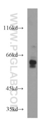
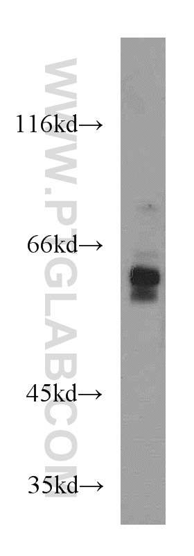
- Jurkat cells were subjected to SDS PAGE followed by western blot with 14833-1-AP(N-PAC antibody) at dilution of 1:500
- Sample type
- cell line
Supportive validation
- Submitted by
- Proteintech Group (provider)
- Main image

- Experimental details
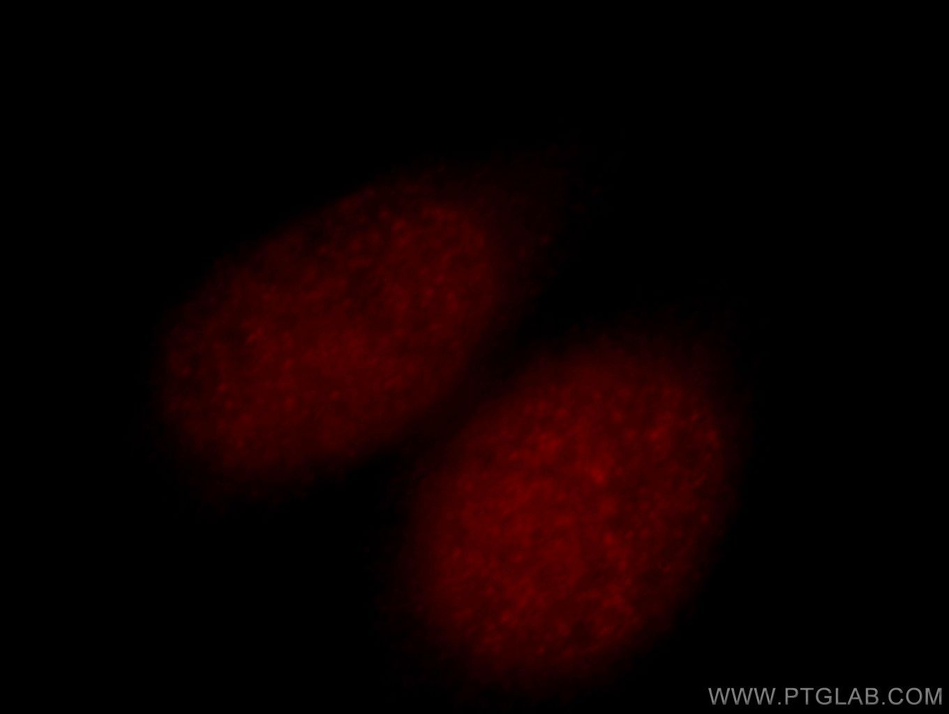
- Immunofluorescent analysis of Hela cells, using N-PAC antibody 14833-1-AP at 1:25 dilution and Rhodamine-labeled goat anti-rabbit IgG (red).
- Sample type
- cell line
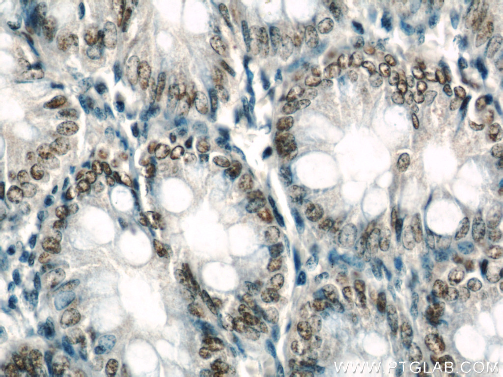
Supportive validation
- Submitted by
- Proteintech Group (provider)
- Main image

- Experimental details
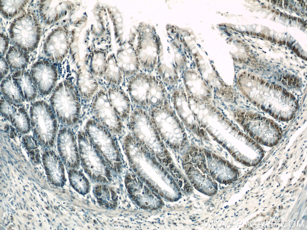
- Immunohistochemical of paraffin-embedded human colon using 14833-1-AP(N-PAC antibody) at dilution of 1:50 (under 40x lens)
- Sample type
- tissue
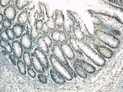
- Submitted by
- Proteintech Group (provider)
- Main image
- Experimental details
- Immunohistochemical of paraffin-embedded human colon using 14833-1-AP(N-PAC antibody) at dilution of 1:50 (under 10x lens)
- Sample type
- tissue