Explore
 Validate
 Learn
Learn Western blot
 Immunohistochemistry
Antibody data
- Antibody Data
- Antigen structure
- References [2]
- Comments [0]
- Validations
- Immunohistochemistry [1]
- Other assay [2]
Submit
Validation data
Reference
Comment
Report error
- Product number
- PA3-070 - Provider product page

- Provider
- Invitrogen Antibodies
- Product name
- SMPX Polyclonal Antibody
- Antibody type
- Polyclonal
- Antigen
- Other
- Description
- PA3-070 detects SMPX from human, mouse, and rat samples. PA3-070 has been successfully used in Western blot, immunocytochemistry, immunohistochemistry (paraffin) procedures. By Western blot, this antibody detects a 9 kDa protein corresponding to mouse SMPX. The PA3-070 antigen is the COOH-terminal amino acids of mouse SMPX (6684) NLSEIQNVKSELKFVPKGE coupled to KLH.
- Reactivity
- Human, Mouse, Rat
- Host
- Rabbit
- Isotype
- IgG
- Vial size
- 100 μL
- Concentration
- Conc. Not Determined
- Storage
- -20°C, Avoid Freeze/Thaw Cycles
Submitted references Missense mutations in small muscle protein X-linked (SMPX) cause distal myopathy with protein inclusions.
The nuclear receptor NOR-1 regulates the small muscle protein, X-linked (SMPX) and myotube differentiation.
Johari M, Sarparanta J, Vihola A, Jonson PH, Savarese M, Jokela M, Torella A, Piluso G, Said E, Vella N, Cauchi M, Magot A, Magri F, Mauri E, Kornblum C, Reimann J, Stojkovic T, Romero NB, Luque H, Huovinen S, Lahermo P, Donner K, Comi GP, Nigro V, Hackman P, Udd B
Acta neuropathologica 2021 Aug;142(2):375-393
Acta neuropathologica 2021 Aug;142(2):375-393
The nuclear receptor NOR-1 regulates the small muscle protein, X-linked (SMPX) and myotube differentiation.
Ferrán B, Martí-Pàmies I, Alonso J, Rodríguez-Calvo R, Aguiló S, Vidal F, Rodríguez C, Martínez-González J
Scientific reports 2016 May 16;6:25944
Scientific reports 2016 May 16;6:25944
No comments: Submit comment
Supportive validation
- Submitted by
- Invitrogen Antibodies (provider)
- Main image
- Experimental details
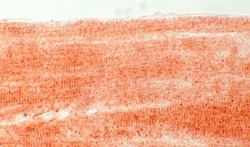
- Immunohistochemistry of SMPX in mouse cardiomyocytes

Supportive validation
- Submitted by
- Invitrogen Antibodies (provider)
- Main image
- Experimental details
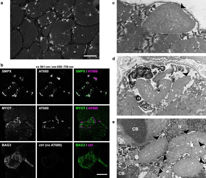
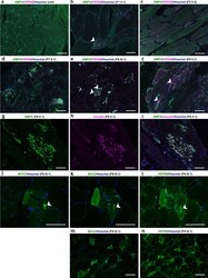
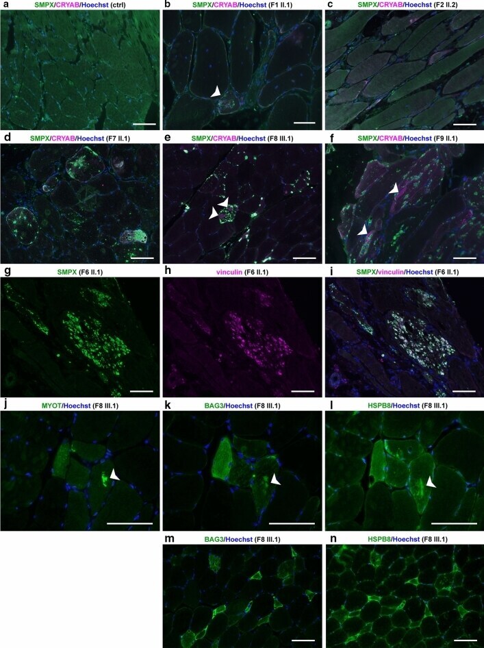
- Fig. 4 Immunofluorescence microscopy. a - f Immunofluorescent double staining; merged images are shown, SMPX is green and CRYAB is magenta . In control skeletal muscle (ctrl) free of neuromuscular disease, SMPX shows diffuse cytoplasmic and focal subsarcolemmal staining pattern. Muscle biopsy of F1 II.1 b shows moderate SMPX accumulation in a single fiber ( white arrowhead ), whereas in patient F2 II.2 c there is no SMPX accumulation. In probands from families F7, F8 and F9 ( d - f ), multiple SMPX-positive sarcoplasmic inclusions are present in several fibers. In F8 III.2 ( e ) and F9 II.1 ( f ), separate myofibrillar CRYAB accumulation is observed ( white arrowheads ), which does not co-localize with SMPX labeling. g - i Double immunostaining of F6 II.1. Single channel and merged images are shown; SMPX is green , vinculin is magenta . SMPX-positive protein inclusions ( g ) are also positive for vinculin ( h ), merged image ( i ). j - l ; serial sections Myofibrillar accumulation ( arrow ) is positive for myotilin ( j ), and also for the CASA proteins BAG3 ( k ) and HSPB8 ( l ). m - n ; serial sections) Several (atrophic) fibers show sarcoplasmic up-regulation of both BAG3 ( m ) and ( n ) HSPB8. Scale bars = 100 um
- Submitted by
- Invitrogen Antibodies (provider)
- Main image

- Experimental details
- Fig. 5 SMPX aggregation in patient muscle. a Congo red staining in fluorescence microscopy using Texas red filter showing abundant positive sarcoplasmic inclusions in muscle biopsy from patient F9 II.1 (scale bar = 50 um). b Confocal sections of a muscle biopsy from patient F8 III.1. Immunostaining of SMPX or myotilin ( green ) combined with Amytracker 680 (AT680, magenta ) shows Amytracker fluorescence in SMPX inclusions. A serial section of the same fiber shows no signal when Amytracker is omitted (BAG3/ctrl), showing that the protein inclusions are not autofluorescent. c - e EM findings of patient F9 II.1. c One large subsarcolemmal protein inclusion with accumulation of filamentous amyloid-like material ( black arrowhead ). d Myeloid bodies ( white arrowhead ) surrounding sarcoplasmic inclusions ( black arrowhead ). e Two classic cytoplasmic bodies (CB) with radiating filaments, together with six individual sarcoplasmic inclusions ( black arrowheads ) and nucleus (N)