 Explore
 Validate
 Learn
Learn Western blot
 ELISA
ELISA Immunocytochemistry
Antibody data
- Antibody Data
- Antigen structure
- References [0]
- Comments [0]
- Validations
- Immunocytochemistry [3]
- Immunohistochemistry [1]
- Flow cytometry [2]
Submit
Validation data
Reference
Comment
Report error
- Product number
- MA5-38467 - Provider product page

- Provider
- Invitrogen Antibodies
- Product name
- GP130 Monoclonal Antibody (8D4D2)
- Antibody type
- Monoclonal
- Antigen
- Purifed from natural sources
- Description
- This antibody has been tested in indirect ELISA.
- Reactivity
- Human
- Host
- Mouse
- Isotype
- IgG
- Antibody clone number
- 8D4D2
- Vial size
- 100 μg
- Concentration
- 1 mg/mL
- Storage
- Store at 4°C short term. For long term storage, store at -20°C, avoiding freeze/thaw cycles.
No comments: Submit comment
Supportive validation
- Submitted by
- Invitrogen Antibodies (provider)
- Main image
- Experimental details
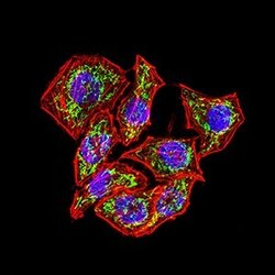
- Immunocytochemistry/Immunofluorescence analysis of GP130 in HeLa cells using GP130 Monoclonal Antibody (Product # MA5-38467) (green). Blue: DRAQ5 fluorescent DNA dye. Red: Actin filaments have been labeled with Alexa Fluor- 555 phalloidin. Secondary antibody from Fisher (Product # 35503)

- Submitted by
- Invitrogen Antibodies (provider)
- Main image
- Experimental details
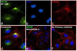
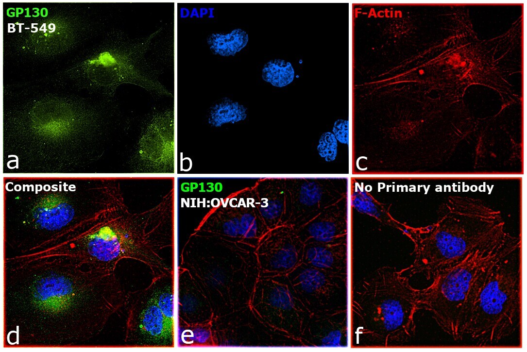
- Immunofluorescence analysis of IL6ST was performed using 70% confluent log phase BT-549 cells. The cells were fixed with 4% paraformaldehyde for 10 minutes, permeabilized with 0.1% Triton™ X-100 for 15 minutes, and blocked with 2% BSA for 1 hour at room temperature. The cells were labeled with GP130 Monoclonal Antibody (8D4D2) (Product # MA5-38467) at 1:200 dilution in 0.1% BSA, incubated at 4 degree celsius overnight and then labeled with Donkey anti-Mouse IgG (H+L) Highly Cross-Adsorbed Secondary Antibody, Alexa Fluor Plus 488 (Product # A32766, 1:2000), for 45 minutes at room temperature (Panel a: Green). Nuclei (Panel b: Blue) were stained with ProLong™ Diamond Antifade Mountant with DAPI (Product # P36962). F-actin (Panel c: Red) was stained with Rhodamine Phalloidin (Product # R415, 1:300). Panel d represents the merged image showing Golgi network and cell membrane localization. Panel e represents the low expression model NIH:OVCAR3 with no signal . Panel f represents control cells with no primary antibody to assess background. The images were captured at 60x magnification.

- Submitted by
- Invitrogen Antibodies (provider)
- Main image

- Experimental details
- Immunofluorescence analysis of IL6ST was performed using 70% confluent log phase BT-549 cells. The cells were fixed with 4% paraformaldehyde for 10 minutes, permeabilized with 0.1% Triton™ X-100 for 15 minutes, and blocked with 2% BSA for 1 hour at room temperature. The cells were labeled with GP130 Monoclonal Antibody (8D4D2) (Product # MA5-38467) at 1:200 dilution in 0.1% BSA, incubated at 4 degree celsius overnight and then labeled with Donkey anti-Mouse IgG (H+L) Highly Cross-Adsorbed Secondary Antibody, Alexa Fluor Plus 488 (Product # A32766, 1:2000), for 45 minutes at room temperature (Panel a: Green). Nuclei (Panel b: Blue) were stained with ProLong™ Diamond Antifade Mountant with DAPI (Product # P36962). F-actin (Panel c: Red) was stained with Rhodamine Phalloidin (Product # R415, 1:300). Panel d represents the merged image showing Golgi network and cell membrane localization. Panel e represents the low expression model NIH:OVCAR3 with no signal . Panel f represents control cells with no primary antibody to assess background. The images were captured at 60x magnification.
Supportive validation
- Submitted by
- Invitrogen Antibodies (provider)
- Main image
- Experimental details
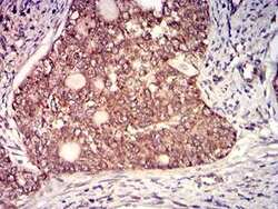
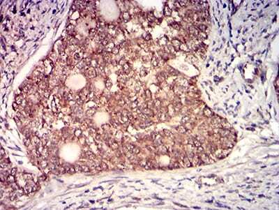
- Immunohistochemistry (Paraffin) analysis of GP130 in paraffin-embedded cervical cancer tissues using GP130 Monoclonal Antibody (Product # MA5-38467) and DAB staining.
Supportive validation
- Submitted by
- Invitrogen Antibodies (provider)
- Main image
- Experimental details
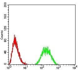
- Flow Cytometry analysis of GP130 in HL-60 cells using GP130 Monoclonal Antibody (Product # MA5-38467) (green) and negative control (red).

- Submitted by
- Invitrogen Antibodies (provider)
- Main image

- Experimental details
- Flow Cytometry analysis of GP130 in HL-60 cells using GP130 Monoclonal Antibody (Product # MA5-38467) (green) and negative control (red).
