 Explore
 Validate
 Learn
LearnPA5-23076
antibody from Invitrogen Antibodies
Targeting: GOLM1
bA379P1.3, C9orf155, FLJ23608, GOLPH2, GP73
 Western blot
Antibody data
- Antibody Data
- Antigen structure
- References [0]
- Comments [0]
- Validations
- Western blot [2]
- Immunocytochemistry [4]
Submit
Validation data
Reference
Comment
Report error
- Product number
- PA5-23076 - Provider product page

- Provider
- Invitrogen Antibodies
- Product name
- GOLPH2 Polyclonal Antibody
- Antibody type
- Polyclonal
- Antigen
- Synthetic peptide
- Description
- PA5-23076 reacts with GOLM1 in human samples. PA5-23076 has been successfully used in Western Blot and Immunocytochemistry/Immunofluorescence applications. The PA5-23076 immunogen is a genomic peptide made to an internal region of the human GOLM1 protein (within residues 250-400).
- Reactivity
- Human, Mouse
- Host
- Rabbit
- Isotype
- IgG
- Vial size
- 100 µL
- Concentration
- 0.61 mg/mL
- Storage
- Store at 4°C short term. For long term storage, store at -20°C, avoiding freeze/thaw cycles.
No comments: Submit comment
Supportive validation
- Submitted by
- Invitrogen Antibodies (provider)
- Main image

- Experimental details
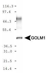
- Western blot analysis of GOLPH2 using a polyclonal antibody (Product # PA5-23076).
- Submitted by
- Invitrogen Antibodies (provider)
- Main image
- Experimental details
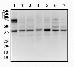
- Western blot analysis of GOLPH2 in whole cell protein from HeLa, 3T3, MEF, Raw264.7, L929, Neuro2A, and PC12. Samples were incubated in GOLPH2 polyclonal antibody (Product # PA5-23076) using a dilution of 0.5 mg/mL followed by an anti-rabbit HRP secondary antibody. Lane 1: Whole cell protein from HeLa; Lane 2: 3T3; Lane 3: MEF; Lane 4: Raw264.7; Lane 5: L929; Lane 6: Neuro2A; Lane 7: PC12 were separated on a 12% gel by SDS-PAGE, transferred to PVDF membrane and blocked in 5% non-fat milk in TBST. Detection: chemiluminescence.

Supportive validation
- Submitted by
- Invitrogen Antibodies (provider)
- Main image

- Experimental details
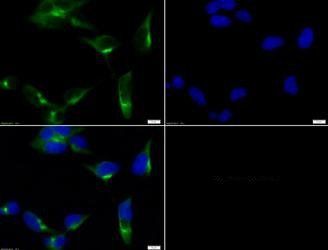
- Immunofluorescent analysis of GOLPH2 using a polyclonal antibody (Product # PA5-23076).
- Submitted by
- Invitrogen Antibodies (provider)
- Main image

- Experimental details
- Immunocytochemistry analysis of GOLPH2 in HeLa cells. Samples were incubated in GOLPH2 polyclonal antibody (Product # PA5-23076) followed by Alexa Fluor 488-conjugated Goat to rabbit IgG secondary antibody (green). Actin filaments were labeled with Alexa Fluor 568 phalloidin (red). DAPI was used to stain the cell nuclei (blue).

- Submitted by
- Invitrogen Antibodies (provider)
- Main image

- Experimental details
- Immunocytochemistry analysis of GOLPH2 in HEK-293 cells. Samples were incubated in GOLPH2 polyclonal antibody (Product # PA5-23076) followed by DyLight 488 (green). Nuclei and alpha-tubulin were counterstained with DAPI (blue) and DyLight 550 (red).
