Explore
 Validate
 Learn
Learn Western blot
 Flow cytometry
Antibody data
- Antibody Data
- Antigen structure
- References [3]
- Comments [0]
- Validations
- Western blot [1]
Submit
Validation data
Reference
Comment
Report error
- Product number
- AF1529 - Provider product page

- Provider
- R&D Systems
- Product name
- Mouse TIM-3 Antibody
- Antibody type
- Polyclonal
- Description
- Antigen Affinity-purified. Detects mouse TIM-3 in direct ELISAs and Western blots. In direct ELISAs, approximately 10% cross-reactivity with recombinant human TIM-3 is observed and less than 5% cross-reactivity with recombinant mouse (rm) TIM-1, rmTIM-2, rmTIM-4, rmTIM-5, rmTIM-6, and rmTIM-7 is observed.
- Reactivity
- Mouse
- Host
- Goat
- Conjugate
- Unconjugated
- Antigen sequence
AAL65156- Isotype
- IgG
- Vial size
- 100 ug
- Concentration
- LYOPH
- Storage
- Use a manual defrost freezer and avoid repeated freeze-thaw cycles. 12 months from date of receipt, -20 to -70 °C as supplied. 1 month, 2 to 8 °C under sterile conditions after reconstitution. 6 months, -20 to -70 °C under sterile conditions after reconstitution.
Submitted references Transcriptomic Hallmarks of Tumor Plasticity and Stromal Interactions in Brain Metastasis.
The HIF-1/glial TIM-3 axis controls inflammation-associated brain damage under hypoxia.
TIM-1 and TIM-3 enhancement of Th2 cytokine production by mast cells.
Wingrove E, Liu ZZ, Patel KD, Arnal-Estapé A, Cai WL, Melnick MA, Politi K, Monteiro C, Zhu L, Valiente M, Kluger HM, Chiang VL, Nguyen DX
Cell reports 2019 Apr 23;27(4):1277-1292.e7
Cell reports 2019 Apr 23;27(4):1277-1292.e7
The HIF-1/glial TIM-3 axis controls inflammation-associated brain damage under hypoxia.
Koh HS, Chang CY, Jeon SB, Yoon HJ, Ahn YH, Kim HS, Kim IH, Jeon SH, Johnson RS, Park EJ
Nature communications 2015 Mar 20;6:6340
Nature communications 2015 Mar 20;6:6340
TIM-1 and TIM-3 enhancement of Th2 cytokine production by mast cells.
Nakae S, Iikura M, Suto H, Akiba H, Umetsu DT, Dekruyff RH, Saito H, Galli SJ
Blood 2007 Oct 1;110(7):2565-8
Blood 2007 Oct 1;110(7):2565-8
No comments: Submit comment
Supportive validation
- Submitted by
- R&D Systems (provider)
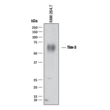
- Main image

- Experimental details
- Detection of Mouse TIM-3 by Western Blot. Western blot shows lysates of RAW 264.7 mouse monocyte/macrophage cell line. PVDF membrane was probed with 2 µg/mL of Goat Anti-Mouse TIM-3 Antigen Affinity-purified Polyclonal Antibody (Catalog # AF1529) followed by HRP-conjugated Anti-Goat IgG Secondary Antibody (Catalog # HAF017). A specific band was detected for TIM-3 at approximately 45-70 kDa (as indicated). This experiment was conducted under reducing conditions and using Immunoblot Buffer Group 1.