Explore
 Validate
 Learn
Learn Western blot
 Flow cytometry
Antibody data
- Antibody Data
- Antigen structure
- References [0]
- Comments [0]
- Validations
- Western blot [1]
- Immunohistochemistry [2]
Submit
Validation data
Reference
Comment
Report error
- Product number
- PA5-46989 - Provider product page

- Provider
- Invitrogen Antibodies
- Product name
- TIM3 Polyclonal Antibody
- Antibody type
- Polyclonal
- Antigen
- Recombinant full-length protein
- Description
- In direct ELISAs, approximately 5% cross-reactivity with recombinant mouse (rm) TIM-3 is observed and less than 1% cross-reactivity with recombinant human TIM-1, rmTIM-1, and rmTIM-2 is observed. Reconstitute at 0.2 mg/mL in sterile PBS.
- Reactivity
- Human
- Host
- Goat
- Isotype
- IgG
- Vial size
- 100 µg
- Concentration
- 0.2 mg/mL
- Storage
- -20° C, Avoid Freeze/Thaw Cycles
No comments: Submit comment
Supportive validation
- Submitted by
- Invitrogen Antibodies (provider)
- Main image
- Experimental details
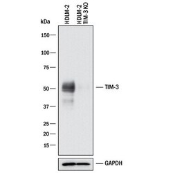
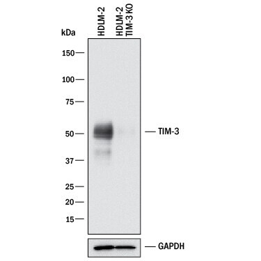
- Knockout validation by Western blot analysis of TIM-3 in lysates of HDLM-2 human Hodgkin’s lymphoma cell line and human TIM-3 knockout HDLM-2 human Hodgkin’s lymphoma cell line (KO). Samples were incubated in TIM-3 polyclonal antibody (Product # PA5-46989) using a dilution of 1 µg/mL followed by a HRP-conjugated Anti-Goat IgG secondary antibody. A specific band was detected for TIM-3 at approximately 50 kDa (as indicated) in the parental HDLM-2 human Hodgkin’s lymphoma cell line, but is not detectable in knockout HDLM-2 human Hodgkin’s lymphoma cell line. GAPDH is shown as a loading control. This experiment was conducted under reducing conditions.
Supportive validation
- Submitted by
- Invitrogen Antibodies (provider)
- Main image
- Experimental details
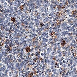
- Immunohistochemical analysis of TIM3 in immersion fixed paraffin-embedded sections of human tonsil. Samples were incubated with TIM3 polyclonal antibody (Product # PA5-46989) using a dilution of 3 µg/mL for 1 hour at room temperature followed by Anti-Goat IgG VisUCyte™ HRP Polymer Antibody. Before incubation with the primary antibody, tissue was subjected to heat-induced epitope retrieval using Antigen Retrieval Reagent-Basic. Tissue was stained using DAB (brown) and counterstained with hematoxylin (blue). Specific staining was localized to cell membranes and extracellular space.

- Submitted by
- Invitrogen Antibodies (provider)
- Main image

- Experimental details
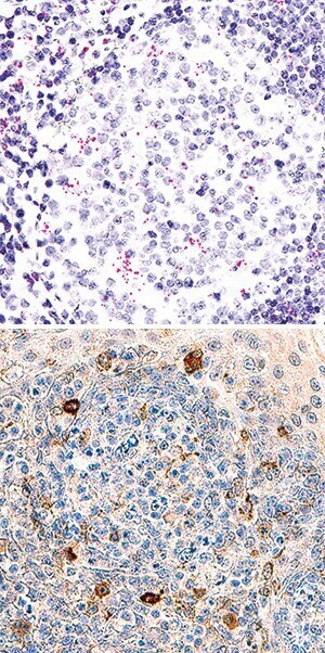
- Immunohistochemical analysis of TIM3 in formalin-fixed paraffin-embedded tissue sections of human tonsil probed with ACD RNAScope® Probe and stained using ACD RNAscope® 2.5 HD Detection Reagents-Red (top image). Samples were incubated in TIM3 polyclonal antibody (Product # PA5-46989) using a dilution of 3 µg/mL for 1 hour at room temperature followed by Anti-Mouse IgG VisUCyte HRP Polymer Antibody and DAB chromogen (lower image, yellow-brown). Tissues were counterstained with hematoxylin (blue).