 Explore
 Validate
 Learn
Learn Western blot
Antibody data
- Antibody Data
- Antigen structure
- References [1]
- Comments [0]
- Validations
- Western blot [1]
- Immunocytochemistry [3]
- Other assay [1]
Submit
Validation data
Reference
Comment
Report error
- Product number
- MA5-24327 - Provider product page

- Provider
- Invitrogen Antibodies
- Product name
- Arylsulfatase B Monoclonal Antibody (708401)
- Antibody type
- Monoclonal
- Antigen
- Recombinant full-length protein
- Description
- Reconstitute in sterile PBS to a final concentration of 0.5 mg/mL.
- Reactivity
- Human
- Host
- Mouse
- Isotype
- IgG
- Antibody clone number
- 708401
- Vial size
- 100 μg
- Concentration
- 0.5 mg/mL
- Storage
- -20°C, Avoid Freeze/Thaw Cycles
Submitted references MiR-29 coordinates age-dependent plasticity brakes in the adult visual cortex.
Napoli D, Lupori L, Mazziotti R, Sagona G, Bagnoli S, Samad M, Sacramento EK, Kirkpartick J, Putignano E, Chen S, Terzibasi Tozzini E, Tognini P, Baldi P, Kwok JC, Cellerino A, Pizzorusso T
EMBO reports 2020 Nov 5;21(11):e50431
EMBO reports 2020 Nov 5;21(11):e50431
No comments: Submit comment
Supportive validation
- Submitted by
- Invitrogen Antibodies (provider)
- Main image
- Experimental details
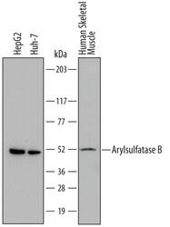
- Western blot analysis of Arylsulfatase B in HepG2 human hepatocellular carcinoma cell line, Huh-7 human hepatoma cell line, and human skeletal muscle tissue. Samples were incubated in Arylsulfatase B monoclonal antibody (Product # MA5-24327) using a dilution of 1 µg/mL followed by a HRP-conjugated Anti-Mouse IgG secondary antibody. A specific band was detected for Arylsulfatase B/ARSB at approximately 47 to 54 kDa (as indicated). This experiment was conducted under reducing conditions.

Supportive validation
- Submitted by
- Invitrogen Antibodies (provider)
- Main image

- Experimental details
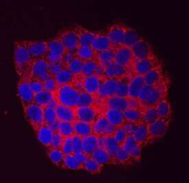
- Immunocytochemistry analysis of Arylsulfatase B in immersion fixed HepG2 human hepatocellular carcinoma cell line. Samples were incubated in Arylsulfatase B monoclonal antibody (Product # MA5-24327) using a dilution of 10 µg/mL for 3 hours at room temperature followed by NorthernLights™ 557-conjugated Anti-Mouse IgG Secondary Antibody (red) and counterstained with DAPI (blue). Specific staining was localized to cytoplasm.
- Submitted by
- Invitrogen Antibodies (provider)
- Main image
- Experimental details
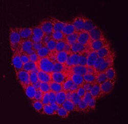
- Immunocytochemistry analysis of Arylsulfatase B in immersion fixed HepG2 human hepatocellular carcinoma cell line. Samples were incubated in Arylsulfatase B monoclonal antibody (Product # MA5-24327) using a dilution of 10 µg/mL for 3 hours at room temperature followed by NorthernLights™ 557-conjugated Anti-Mouse IgG Secondary Antibody (red) and counterstained with DAPI (blue). Specific staining was localized to cytoplasm.
- Submitted by
- Invitrogen Antibodies (provider)
- Main image

- Experimental details
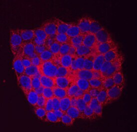
- Immunocytochemistry analysis of Arylsulfatase B in immersion fixed HepG2 human hepatocellular carcinoma cell line. Samples were incubated in Arylsulfatase B monoclonal antibody (Product # MA5-24327) using a dilution of 10 µg/mL for 3 hours at room temperature followed by NorthernLights™ 557-conjugated Anti-Mouse IgG Secondary Antibody (red) and counterstained with DAPI (blue). Specific staining was localized to cytoplasm.

Supportive validation
- Submitted by
- Invitrogen Antibodies (provider)
- Main image
- Experimental details
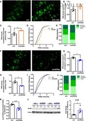
- 6 Figure Manipulations of miR-29a levels promote changes in PNN s A Images of WFA-labeled PNNs of scrambled-treated visual cortex (left) and miR-29a mimic (right) in young mice. Scale bar 60 mum. B, C Quantification of PNN density (scr vs. mim29a, N = 3 mice per group; t -test, P = 0.20) and intensity for each animal (scr vs. mim29a, N = 3 mice per group; t -test, P = 0.014). D Cumulative frequency of PNN staining intensity in scrambled- and mimic-treated young mice scr ( N = 1,438 cells vs. mim29a N = 1,749 cells, Kolmogorov-Smirnov test, P < 0.0001). E PNN distribution in classes of intensity in mice treated with scrambled and mimic 29a. F Images of WFA-labeled PNNs of scrambled-treated visual cortex (left) and amiR-29a LNA (right) in adult mice. Scale bar 60 mum. G, H Quantification of PNN density (scr vs. amiR-29a, N = 3 mice per group; t -test, P = 0.03) and intensity for each animal (scr vs. amiR-29a, N = 3 mice per group; t -test, P = 0.047). I Cumulative frequency of PNN intensity in scrambled and amiR-29a-treated adult mice. scr N = 1,883 cells vs. amiR-29a N = 1,937 cells, Kolmogorov-Smirnov test, P < 0.0001. J PNN distribution in classes of intensity in mice treated with scrambled and amiR-29a. K Biochemical quantification of C4S in adult amiR-29a-treated mice ( N = 5) as compared to scrambled animals ( N = 4). t -test, P = 0.0008. L Example of arylsulfatase B (ArsB) Western blot. Left: The lanes corresponding to the treated (treat scr or treat amiR-29a) and untrea
