Explore
 Validate
 Learn
Learn Western blot
 ELISA
ELISA Immunocytochemistry
Antibody data
- Antibody Data
- Antigen structure
- References [2]
- Comments [0]
- Validations
- Immunocytochemistry [3]
- Immunohistochemistry [2]
- Other assay [6]
Submit
Validation data
Reference
Comment
Report error
- Product number
- PA5-113283 - Provider product page

- Provider
- Invitrogen Antibodies
- Product name
- MRAP2 Polyclonal Antibody
- Antibody type
- Polyclonal
- Antigen
- Recombinant full-length protein
- Reactivity
- Human, Mouse
- Host
- Rabbit
- Isotype
- IgG
- Vial size
- 100 μg
- Concentration
- 3.5 mg/mL
- Storage
- -20°C or -80°C if preferred
Submitted references Identification of Regions Involved in the Physical Interaction between Melanocortin Receptor Accessory Protein 2 and Prokineticin Receptor 2.
Arginine 125 Is an Essential Residue for the Function of MRAP2.
Fullone MR, Maftei D, Vincenzi M, Lattanzi R, Miele R
Biomolecules 2022 Mar 20;12(3)
Biomolecules 2022 Mar 20;12(3)
Arginine 125 Is an Essential Residue for the Function of MRAP2.
Fullone MR, Maftei D, Vincenzi M, Lattanzi R, Miele R
International journal of molecular sciences 2022 Aug 30;23(17)
International journal of molecular sciences 2022 Aug 30;23(17)
No comments: Submit comment
Supportive validation
- Submitted by
- Invitrogen Antibodies (provider)
- Main image
- Experimental details
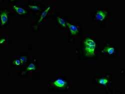
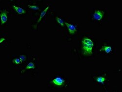
- Immunocytochemichal analysis of MRAP2 in U251 cells using a Polyclonal antibody (Product # PA5-113283) at a dilution of 1:100. An Alexa Fluor 488-congugated Goat Anti-Rabbit IgG(H+L) secondary antibdoy was used.
- Submitted by
- Invitrogen Antibodies (provider)
- Main image

- Experimental details
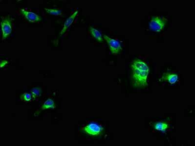
- Immunocytochemichal analysis of MRAP2 in U251 cells using a Polyclonal antibody (Product # PA5-113283) at a dilution of 1:100. An Alexa Fluor 488-congugated Goat Anti-Rabbit IgG(H+L) secondary antibdoy was used.
- Submitted by
- Invitrogen Antibodies (provider)
- Main image
- Experimental details
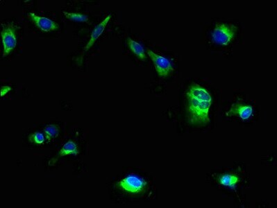
- Immunocytochemichal analysis of MRAP2 in U251 cells using a Polyclonal antibody (Product # PA5-113283) at a dilution of 1:100. An Alexa Fluor 488-congugated Goat Anti-Rabbit IgG(H+L) secondary antibdoy was used.

Supportive validation
- Submitted by
- Invitrogen Antibodies (provider)
- Main image

- Experimental details
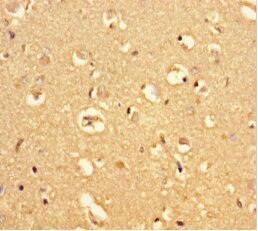
- Immunocytochemical analysis of paraffin-embeded MRAP2 in human brain tissue using a MRAP2 Polyclonal antibody (Product # PA5-113283) at a dilution of 1:100.
- Submitted by
- Invitrogen Antibodies (provider)
- Main image
- Experimental details
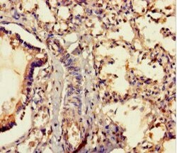
- Immunocytochemical analysis of paraffin-embeded MRAP2 in human lung tissue using a MRAP2 Polyclonal antibody (Product # PA5-113283) at a dilution of 1:100.

Supportive validation
- Submitted by
- Invitrogen Antibodies (provider)
- Main image

- Experimental details
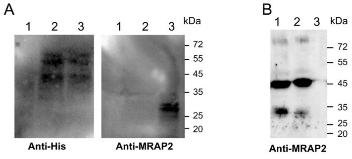
- Interaction of MRAP2 with the N-terminal region of PKR2. ( A ) Membranes from yeast co-expressing MRAP2 with PKR2 or DeltaN-PKR2 were co-precipitated using Ni-NTA His-bind resin and were resolved by 12% SDS-PAGE. The immunoblots were probed with anti-His (left panel) and anti-MRAP2 (right panel) antibodies. Lane 1: Cy12946, lane 2: DeltaN-PKR2, and lane 3: PKR2. ( B ) The GST fusion protein PKR2-NT was used to pull-down MRAP2, and the elution solutions obtained were resolved 15% SDS-PAGE analyzed by Western blotting using an anti-MRAP2 antibody. Lane 1: input; lane 2: PKR2-NT eluate; and lane 3: GST eluate.
- Submitted by
- Invitrogen Antibodies (provider)
- Main image

- Experimental details
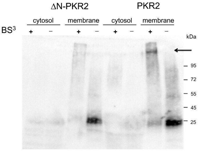
- Cross-linking of MRAP2 with PKR2 or DeltaN-PKR2 receptors. Membrane and cytosolic proteins prepared from S. cerevisiae cells expressing the PKR2-MRAP2 and DeltaN-PKR2 -MRAP2 were incubated in the presence (+) or absence (-) of BS 3 . Proteins were immunoblotted and probed with an anti-MRAP2 antibody. The arrow indicates the complex PKR2/MRAP2.
- Submitted by
- Invitrogen Antibodies (provider)
- Main image
- Experimental details
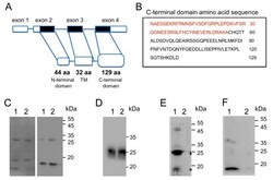
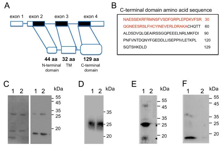
- Analysis of MRAP2 regions that bind PKR2. ( A ) Scheme of human MRAP2 gene. Exon coding sequences are indicated as black bars and the untranslated sequences are shown as white bars. ( B ) In the box, the amino acid sequence of the C-terminal MRAP2 domain; the sequence present in the 131CT-MRAP2 domain is indicated in red. ( C ) SDS PAGE and Western blot analysis of CT-MRAP2 domain expression in E. coli ; lane 1: 5 mug; lane 2: 10 mug. ( D ) Western blot analysis of native PAGE of CT-MRAP2 domain expression in E. coli ; lane 1: 10 mug; lane 2: 15 mug. ( E ) The GST fusion protein and PKR2-NT were used to pull-down the CT-MRAP2 domain. Solutions obtained by elution were analyzed by Western blotting: lane 1; PKR2-NT eluate; lane 2: GST eluate. ( F ) The GST fusion protein and PKR2-NT were used to pull-down the 131CT-MRAP2 domain. Solutions obtained by elution were analyzed by Western blotting: lane 1; PKR2-NT eluate; lane 2: GST eluate.
- Submitted by
- Invitrogen Antibodies (provider)
- Main image
- Experimental details
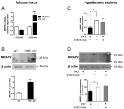
- Figure 5 MRAP2 expression in adipocytes and hypothalamic explants. MRAP2 levels in adipose tissue were analyzed by ( A ) real-time RT-PCR and ( B ) Western blot analysis. MRAP2 levels in hypothalamus explants analyzed by ( C ) real-time RT-PCR and ( D ) Western blot analysis (arrow). Data are expressed as the mean +- SEM of three separate experiments (n = 4 per group). Statistical analyses were performed using Student's t -test where ** p < 0.01; *** p < 0.001 vs. WT ( A , B ) and one-way ANOVA followed by Tukey's post-test, * p < 0.05 ( C , D ).

- Submitted by
- Invitrogen Antibodies (provider)
- Main image
- Experimental details
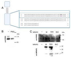
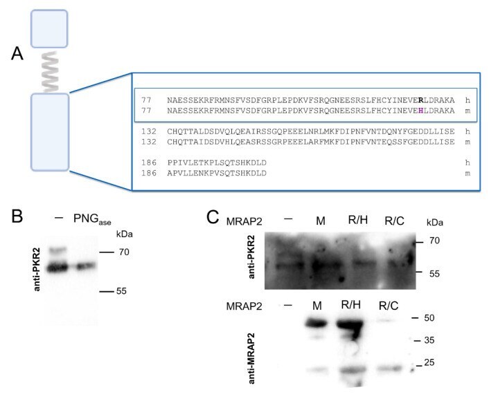
- Analysis of PKR2 glycosylation in CHO cells in the presence of mouse MRAP2 and MRAP2 mutants. ( A ) Alignment of the human and mouse MRAP2 C-terminal domain. The sequence alignments of the region important for dimerization are shown in the box. ( B ) CHO stably expressing PKR2 untreated membrane proteins or those treated with PNGase F were analyzed by western blot analysis using an anti-PKR2 antibody. ( C ) Membrane proteins CHO stably expressing PKR2 were analyzed by western blot analysis using anti-PKR2 and anti-MRAP2 antibodies. In the absence of MRAP2, -; in the presence of mouse MRAP2, M; MRAP2 R125H mutant, R/H; MRAP2 R125C mutant, R/C.
- Submitted by
- Invitrogen Antibodies (provider)
- Main image
- Experimental details
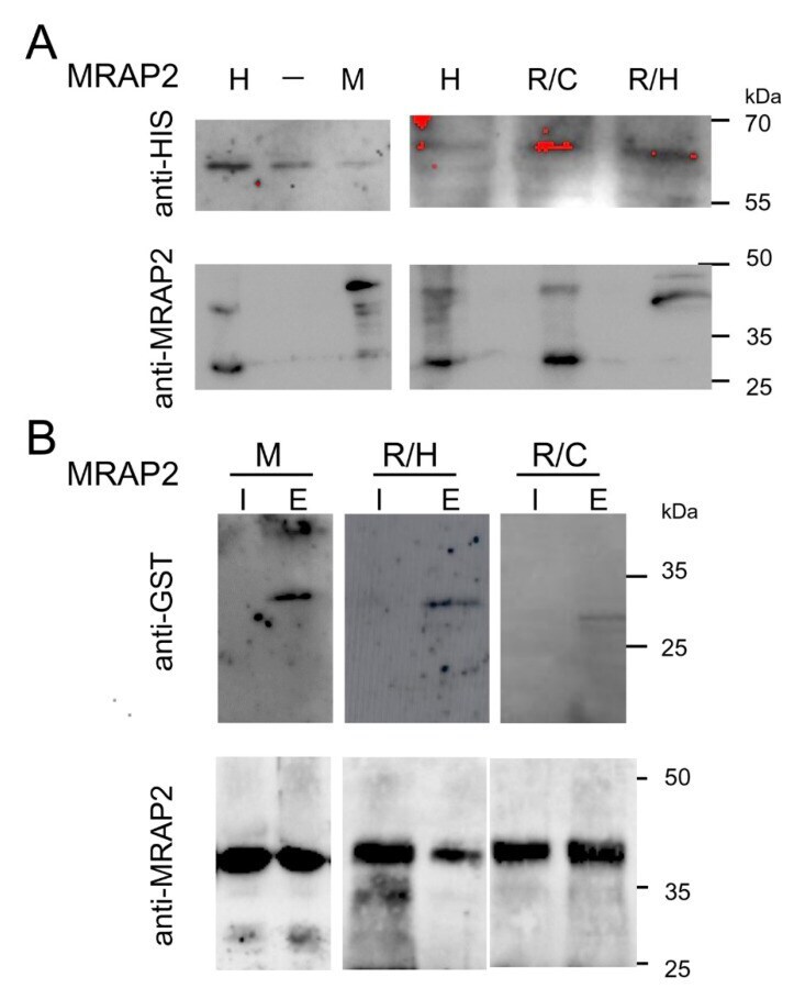
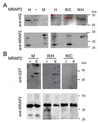
- Interaction of MRAP2 isoforms with PKR2. ( A ) Membrane proteins from yeast co-expressing MRAP2 isoforms with PKR2 were co-precipitated using Ni-NTA His-bind resin and resolved by 12% SDS-PAGE. The immunoblots were probed with anti-His and anti-MRAP2 antibodies. Mouse MRAP2, M; human MRAP2, H; MRAP2 R125H, R/H; R125C MRAP2, R/C. ( B ) GST pull-down experiments. The GST fusion protein of PKR2-NT was used to pull down MRAP2 isoforms. Mouse MRAP2, M; R125H mutant, R/H; R125C mutant, R/C. Input and solutions obtained by elution were analyzed by western blotting with anti-GST and anti-MRAP2. I, input; E, eluate.