Explore
 Validate
 Learn
Learn Western blot
 Immunohistochemistry
Antibody data
- Antibody Data
- Antigen structure
- References [1]
- Comments [0]
- Validations
- Immunohistochemistry [2]
- Other assay [2]
Submit
Validation data
Reference
Comment
Report error
- Product number
- PA5-20589 - Provider product page

- Provider
- Invitrogen Antibodies
- Product name
- POLR3F Polyclonal Antibody
- Antibody type
- Polyclonal
- Antigen
- Synthetic peptide
- Description
- A suggested positive control is human brain tissue lysate. PA5-20589 can be used with blocking peptide PEP-0709.
- Reactivity
- Human, Mouse, Rat
- Host
- Rabbit
- Isotype
- IgG
- Vial size
- 100 μg
- Concentration
- 1 mg/mL
- Storage
- Maintain refrigerated at 2-8°C for up to 3 months. For long term storage store at -20°C
Submitted references Truncated PARP1 mediates ADP-ribosylation of RNA polymerase III for apoptosis.
Chen Q, Ma K, Liu X, Chen SH, Li P, Yu Y, Leung AKL, Yu X
Cell discovery 2022 Jan 18;8(1):3
Cell discovery 2022 Jan 18;8(1):3
No comments: Submit comment
Supportive validation
- Submitted by
- Invitrogen Antibodies (provider)
- Main image
- Experimental details
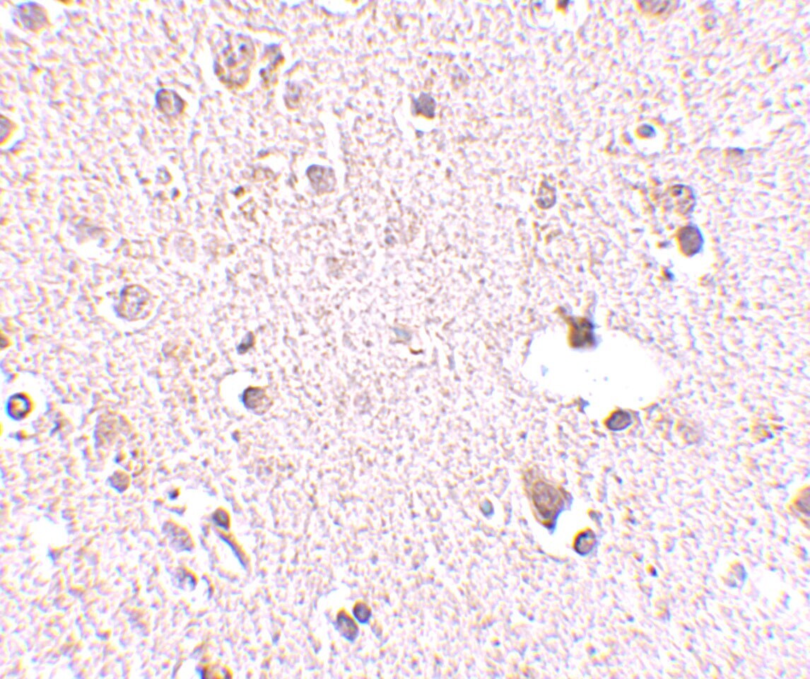
- Immunohistochemistry of POLR3F in human brain tissue with POLR3F Polyclonal Antibody (Product # PA5-20589) at 2.5 µg/mL.
- Submitted by
- Invitrogen Antibodies (provider)
- Main image
- Experimental details
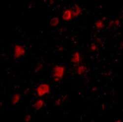
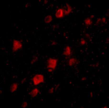
- Immunofluorescence of POLR3F in human brain tissue with POLR3F Polyclonal Antibody (Product # PA5-20589) at 20 µg/mL.
Supportive validation
- Submitted by
- Invitrogen Antibodies (provider)
- Main image
- Experimental details
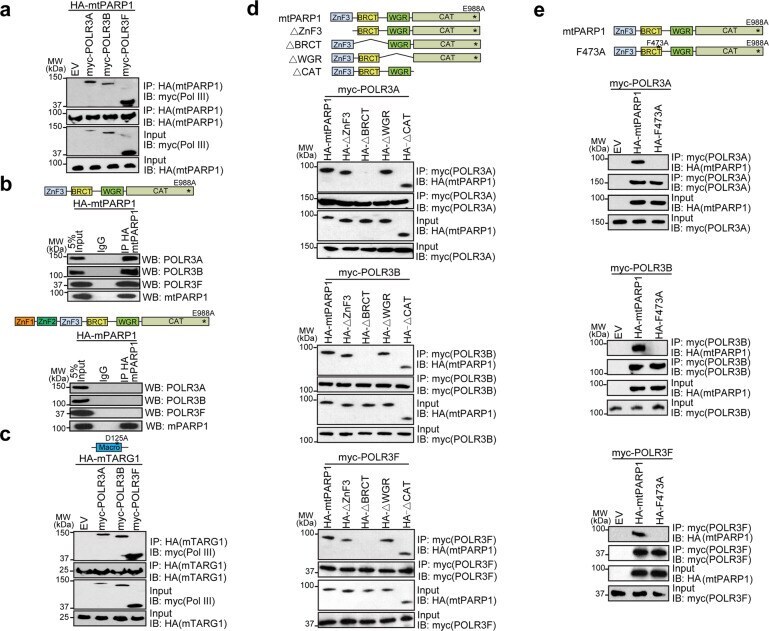
- Fig. 2 The BRCT domain of tPARP1 interacts with the Pol III complex. a U2OS-PARP1 knockout cells were transfected with the myc-empty vector (EV), myc-POLR3A, myc-POLR3B, or myc-POLR3F together with HA-mtPARP1, followed by IP and western blotting with the indicated antibodies. b mtPARP1 or full-length mPARP1 was ectopically expressed in U2OS-PARP1 knockout cells. IP was performed with IgG as a negative control. c U2OS-PARP1 knockout cells were transfected with the myc-empty vector (EV), myc-POLR3A, myc-POLR3B, or myc-POLR3F together with HA-mTARG1, followed by IP and western blotting with the indicated antibodies. d U2OS-PARP1 knockout cells were transfected with the HA-tagged truncation mutants of mtPARP1 together with myc-POLR3A (upper panel), myc-POLR3B (middle panel), or myc-POLR3F (lower panel). e U2OS-PARP1 knockout cells were transfected with the indicated combinations of HA-tagged mtPARP1 or mtPARP1 F473A mutant together with myc-POLR3A (upper panel), myc-POLR3B (middle panel), or myc-POLR3F (lower panel).
- Submitted by
- Invitrogen Antibodies (provider)
- Main image

- Experimental details
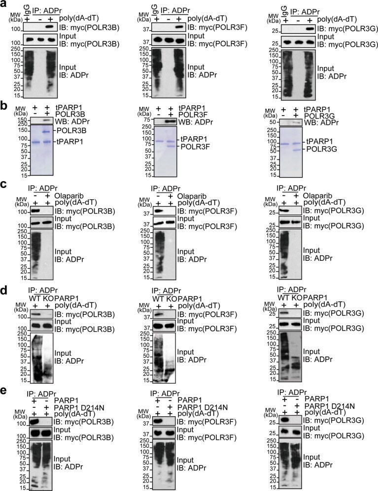
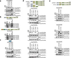
- Fig. 5 RNA Pol III subunits are substrates of tPARP1. a POLR3B, POLR3F, and POLR3G are identified as substrates of tPARP1. Individual subunits of Pol III with myc tag was expressed in U2OS cells, followed by poly(dA-dT)-stimulated apoptosis. Anti-ADPr antibody was used for IP; and the complex was analyzed by western blotting with anti-myc antibody. b POLR3B, POLR3F, and POLR3G are substrates of tPARP1 in vitro. Auto-ADP-ribosylation was detected by anti-ADPr antibody (upper panel). Recombinant protein in each reaction was also examined by the SDS-PAGE with Coomassie blue staining (lower panel). c ADP-ribosylation of POLR3B, POLR3F, or POLR3G was examined in U2OS cells treated with or without 1 uM olaparib for 1 h. d ADP-ribosylation of POLR3B, POLR3F, or POLR3G was examined in wild-type and PARP1 knockout U2OS cells. e ADP-ribosylation of POLR3B, POLR3F, or POLR3G was examined in U2OS cells expressing wild-type or caspase 3-resistant (D214N) PARP1. Note that the cells were harvested after 6-h treatment with 5 mug/mL poly(dA-dT) in a , c - e .