Explore
 Validate
 Learn
LearnPA5-24208
antibody from Invitrogen Antibodies
Targeting: DHTKD1
CMT2Q, DKFZP762M115, KIAA1630, MGC3090
 Western blot
 Other assay
Antibody data
- Antibody Data
- Antigen structure
- References [2]
- Comments [0]
- Validations
- Other assay [2]
Submit
Validation data
Reference
Comment
Report error
- Product number
- PA5-24208 - Provider product page

- Provider
- Invitrogen Antibodies
- Product name
- DHTKD1 Polyclonal Antibody
- Antibody type
- Polyclonal
- Antigen
- Synthetic peptide
- Description
- This antibody is predicted to react with rat based on sequence homology.
- Reactivity
- Human, Mouse
- Host
- Rabbit
- Isotype
- IgG
- Vial size
- 400 μL
- Concentration
- 0.5 mg/mL
- Storage
- Store at 4°C short term. For long term storage, store at -20°C, avoiding freeze/thaw cycles.
Submitted references Synthetic analogues of 2-oxo acids discriminate metabolic contribution of the 2-oxoglutarate and 2-oxoadipate dehydrogenases in mammalian cells and tissues.
Dual mode action of mangiferin in mouse liver under high fat diet.
Artiukhov AV, Grabarska A, Gumbarewicz E, Aleshin VA, Kähne T, Obata T, Kazantsev AV, Lukashev NV, Stepulak A, Fernie AR, Bunik VI
Scientific reports 2020 Feb 5;10(1):1886
Scientific reports 2020 Feb 5;10(1):1886
Dual mode action of mangiferin in mouse liver under high fat diet.
Lim J, Liu Z, Apontes P, Feng D, Pessin JE, Sauve AA, Angeletti RH, Chi Y
PloS one 2014;9(3):e90137
PloS one 2014;9(3):e90137
No comments: Submit comment
Supportive validation
- Submitted by
- Invitrogen Antibodies (provider)
- Main image
- Experimental details
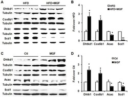
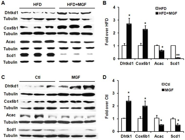
- Figure 6 MGF induces Dhtkd1 and Cox6b, and suppresses Acac and Scd1 in hepatic tissues and cells. (A) Western blots of Dhtkd1, Cox6b1, Acac and Scd1 in proteins extracted from liver tissues from C57BL6/J mice fed with HFD +- MGF for 18 weeks. (B) Quantification of protein expression in (A). Liver tissues were from 2 groups (HFD +- MGF), 3 mice per group. Values are average +- SEM (n = 3). (C) Western blots of Dhtkd1, Cox6b1, Acac and Scd1 in proteins extracted from mouse primary hepatocytes and treated with or without 200 uM MGF for 24 hours. (D) Quantification of protein expression in (C). Values are average +- SEM (n = 3). *p
- Submitted by
- Invitrogen Antibodies (provider)
- Main image

- Experimental details
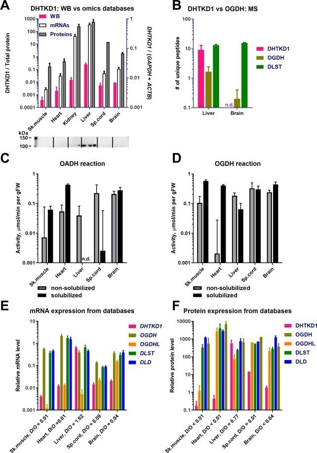
- Figure 2 Levels of the proteins, mRNA and activities of OADHC and OGDHC in different rat tissues. ( A ) Comparison of DHTKD1 expression, determined by Western blot (WB) in our experiments, and extracted from transcriptomics and proteomics databases. The tissue homogenates (n = 3 for each tissue) reacted with anti-DHTKD1 antibodies (Invitrogen). Pink bars correspond to the intensities of bands in 100-110 kDa area (Fig. S1A), which were normalized to the total protein in the lane, measured by 2,2,2-trichloroethanol staining (Fig. S1B). The data extraction from the gene expression databases is described in Materials and Methods. ( B ) Mass-spectrometric detection of the DHTKD1, OGDH, and DLST proteins in mitochondria isolated from the rat liver (n = 6) or cerebral cortex (n = 5). Numbers of unique peptides, detected after in-gel trypsin digestion, are presented. (C,D) Specific activities of the rat tissue homogenates in catalysing the OADH ( C ) and OGDH reactions ( D ), assayed before (grey bars) or after solubilization of membranous compartments by sonication and detergents (black bars). ( E ) Relative levels of mRNAs for the multienzyme complex components DHTKD1 , OGDH , OGDHL , DLST and DLD in the rat tissues. The transcriptomic data are extracted from >=18 independent series of experiments for each tissue. ( F ) Relative levels of the DHTKD1, OGDH, OGDHL, DLST and DLD proteins in the mouse tissues. Except for spinal cord (1 experiment), the protein abundances were extracted