 Explore
 Validate
 Learn
Learn Western blot
Antibody data
- Antibody Data
- Antigen structure
- References [1]
- Comments [0]
- Validations
- Western blot [2]
- Immunocytochemistry [2]
- Immunohistochemistry [1]
Submit
Validation data
Reference
Comment
Report error
- Product number
- MAB7934 - Provider product page

- Provider
- R&D Systems
- Product name
- Human/Mouse/Rat Dihydrofolate Reductase/DHFR Antibody
- Antibody type
- Monoclonal
- Description
- Protein A or G purified from hybridoma culture supernatant. Detects human Dihydrofolate Reductase/DHFR in ELISA. Detects human, mouse and rat Dihydrofolate Reductase in Western Blots.
- Reactivity
- Human, Mouse, Rat
- Host
- Mouse
- Conjugate
- Unconjugated
- Antigen sequence
P00374- Isotype
- IgG
- Antibody clone number
- 872442
- Vial size
- 100 ug
- Concentration
- LYOPH
- Storage
- Use a manual defrost freezer and avoid repeated freeze-thaw cycles. 12 months from date of receipt, -20 to -70 °C as supplied. 1 month, 2 to 8 °C under sterile conditions after reconstitution. 6 months, -20 to -70 °C under sterile conditions after reconstitution.
Submitted references Cross Talk between One-Carbon Metabolism, Eph Signaling, and Histone Methylation Promotes Neural Stem Cell Differentiation.
Fawal MA, Jungas T, Kischel A, Audouard C, Iacovoni JS, Davy A
Cell reports 2018 Jun 5;23(10):2864-2873.e7
Cell reports 2018 Jun 5;23(10):2864-2873.e7
No comments: Submit comment
Supportive validation
- Submitted by
- R&D Systems (provider)
- Main image
- Experimental details
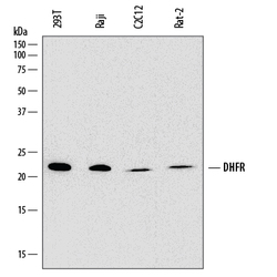
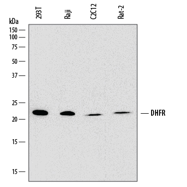
- Detection of Human, Mouse, and Rat Dihydrofolate Reductase/DHFR by Western Blot. Western blot shows lysates of 293T human embryonic kidney cell line, Raji human Burkitt's lymphoma cell line, C2C12 mouse myoblast cell line, Rat-2 rat embryonic fibroblast cell line. PVDF membrane was probed with 1 µg/mL of Mouse Anti-Human Dihydrofolate Reductase/DHFR Monoclonal Antibody (Catalog # MAB7934) followed by HRP-conjugated Anti-Mouse IgG Secondary Antibody (Catalog # HAF018). A specific band was detected for Dihydrofolate Reductase/DHFR at approximately 21 kDa (as indicated). This experiment was conducted under reducing conditions and using Immunoblot Buffer Group 1.
- Submitted by
- R&D Systems (provider)
- Main image
- Experimental details
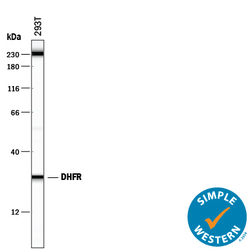
- Detection of Human Dihydrofolate Reductase/DHFR by Simple WesternTM. Simple Western lane view shows lysates of 293T human embryonic kidney cell line, loaded at 0.5 mg/mL. A specific band was detected for Dihydrofolate Reductase/DHFR at approximately 28 kDa (as indicated) using 10 µg/mL of Mouse Anti-Human/Mouse/Rat Dihydrofolate Reductase/DHFR Monoclonal Antibody (Catalog # MAB7934). This experiment was conducted under reducing conditions and using the 12-230 kDa separation system. Non-specific interaction with the 230 kDa Simple Western standard may be seen with this antibody.

Supportive validation
- Submitted by
- R&D Systems (provider)
- Main image

- Experimental details
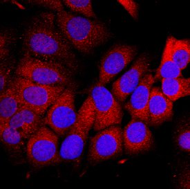
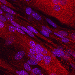
- Dihydrofolate Reductase/DHFR in MCF-7 Human Cell Line. Dihydrofolate Reductase/DHFR was detected in immersion fixed MCF-7 human breast cancer cell line using Mouse Anti-Human Dihydrofolate Reductase/DHFR Monoclonal Antibody (Catalog # MAB7934) at 10 µg/mL for 3 hours at room temperature. Cells were stained using the NorthernLights™ 557-conjugated Anti-Mouse IgG Secondary Antibody (red; Catalog # NL007) and counterstained with DAPI (blue). Specific staining was localized to the cytoplasm. View our protocol for Fluorescent ICC Staining of Cells on Coverslips.
- Submitted by
- R&D Systems (provider)
- Main image
- Experimental details
- Dihydrofolate Reductase/DHFR in C2C12 Mouse Cell Line. Dihydrofolate Reductase/DHFR was detected in immersion fixed C2C12 mouse myoblast cell line using Mouse Anti-Human Dihydrofolate Reductase/DHFR Monoclonal Antibody (Catalog # MAB7934) at 10 µg/mL for 3 hours at room temperature. Cells were stained using the NorthernLights™ 557-conjugated Anti-Mouse IgG Secondary Antibody (red; Catalog # NL007) and counterstained with DAPI (blue). Specific staining was localized to cytoplasm. View our protocol for Fluorescent ICC Staining of Cells on Coverslips.

Supportive validation
- Submitted by
- R&D Systems (provider)
- Main image
- Experimental details
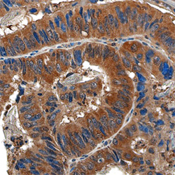
- Dihydrofolate Reductase/DHFR in Human Colon. Dihydrofolate Reductase/DHFR was detected in formalin fixed paraffin-embedded sections of human colon tissue using Mouse Anti-Human Dihydrofolate Reductase/DHFR Monoclonal Antibody (Catalog # MAB7934) at 15 µg/mL overnight at 4 °C. Tissue was stained using the Anti-Mouse HRP-DAB Cell & Tissue Staining Kit (brown; Catalog # CTS002) and counterstained with hematoxylin (blue). Specific staining was localized to the cytoplasm. View our protocol for Chromogenic IHC Staining of Paraffin-embedded Tissue Sections.
