 Explore
 Validate
 Learn
Learn Western blot
 Immunoprecipitation
Antibody data
- Antibody Data
- Antigen structure
- References [0]
- Comments [0]
- Validations
- Immunoprecipitation [1]
- Immunohistochemistry [2]
- Other assay [1]
Submit
Validation data
Reference
Comment
Report error
- Product number
- MA5-38337 - Provider product page

- Provider
- Invitrogen Antibodies
- Product name
- DHFR Recombinant Rabbit Monoclonal Antibody (9B2)
- Antibody type
- Monoclonal
- Antigen
- Synthetic peptide
- Description
- This antibody has been tested in direct-ELISA
- Reactivity
- Human, Mouse, Rat
- Host
- Rabbit
- Isotype
- IgG
- Antibody clone number
- 9B2
- Vial size
- 100 μL
- Concentration
- 0.4 mg/mL
- Storage
- -20°C or -80°C if preferred
No comments: Submit comment
Supportive validation
- Submitted by
- Invitrogen Antibodies (provider)
- Main image
- Experimental details
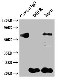
- Immunoprecipitating DHFR in Hela whole cell lysate. Lane 1: Rabbit control IgG Hela whole cell lysate. For western blotting, a HRP-conjugated Protein G antibody was used as the secondary antibody (1:2,000). Lane 2: DHFR Monoclonal Antibody (Product # MA5-38337) (2 µg) + Hela whole cell lysate (500 µg). Lane 3: Hela whole cell lysate (10 µg).

Supportive validation
- Submitted by
- Invitrogen Antibodies (provider)
- Main image
- Experimental details
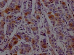
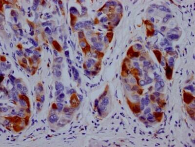
- Immunohistochemistry (Paraffin) analysis of DHFR in paraffin-embedded human breast cancer using DHFR Monoclonal Antibody (Product # MA5-38337) diluted at 1:100. After dewaxing and hydration, antigen retrieval was mediated by high pressure in a citrate buffer (pH 6.0). Section was blocked with 10% normal goat serum 30min at RT. Then primary antibody (1% BSA) was incubated at 4°C overnight. The primary is detected by a Goat anti-rabbit IgG polymer labeled by HRP and visualized using 0.05% DAB.
- Submitted by
- Invitrogen Antibodies (provider)
- Main image

- Experimental details
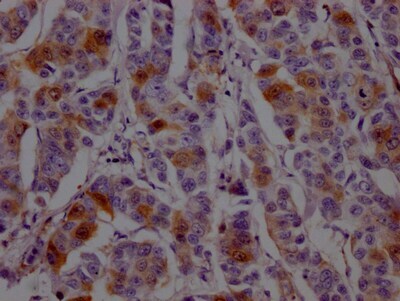
- Immunohistochemistry (Paraffin) analysis of DHFR in paraffin-embedded human liver cancer using DHFR Monoclonal Antibody (Product # MA5-38337) diluted at 1:100. After dewaxing and hydration, antigen retrieval was mediated by high pressure in a citrate buffer (pH 6.0). Section was blocked with 10% normal goat serum 30min at RT. Then primary antibody (1% BSA) was incubated at 4°C overnight. The primary is detected by a Goat anti-rabbit IgG polymer labeled by HRP and visualized using 0.05% DAB.
Supportive validation
- Submitted by
- Invitrogen Antibodies (provider)
- Main image

- Experimental details
- Immunoprecipitating DHFR in Hela whole cell lysate. Lane 1: Rabbit control IgG Hela whole cell lysate. For western blotting, a HRP-conjugated Protein G antibody was used as the secondary antibody (1:2,000). Lane 2: DHFR Monoclonal Antibody (Product # MA5-38337) (2 µg) + Hela whole cell lysate (500 µg). Lane 3: Hela whole cell lysate (10 µg).
