Explore
 Validate
 Learn
Learn Western blot
 Immunocytochemistry
Antibody data
- Antibody Data
- Antigen structure
- References [1]
- Comments [0]
- Validations
- Immunocytochemistry [5]
- Immunohistochemistry [2]
- Other assay [1]
Submit
Validation data
Reference
Comment
Report error
- Product number
- PA5-72964 - Provider product page

- Provider
- Invitrogen Antibodies
- Product name
- CHD7 Polyclonal Antibody
- Antibody type
- Polyclonal
- Antigen
- Synthetic peptide
- Description
- Immunogen has 85% identity to chicken Chd7. Prior to immunostaining paraffin tissues, antigen retrieval with sodium citrate buffer (pH 6.0) is recommended.
- Reactivity
- Human, Mouse
- Host
- Rabbit
- Isotype
- IgG
- Vial size
- 100 μL
- Concentration
- 1 mg/mL
- Storage
- Store at 4°C short term. For long term storage, store at -20°C, avoiding freeze/thaw cycles.
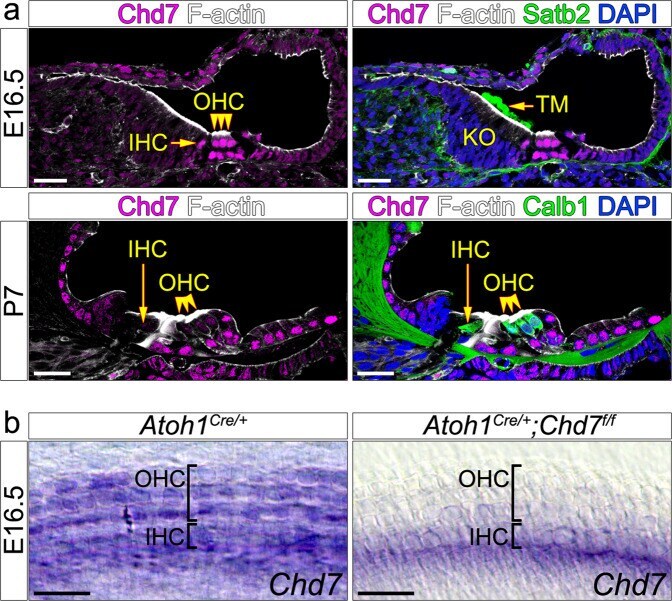
Submitted references The chromatin remodelling factor Chd7 protects auditory neurons and sensory hair cells from stress-induced degeneration.
Ahmed M, Moon R, Prajapati RS, James E, Basson MA, Streit A
Communications biology 2021 Nov 3;4(1):1260
Communications biology 2021 Nov 3;4(1):1260
No comments: Submit comment
Supportive validation
- Submitted by
- Invitrogen Antibodies (provider)
- Main image
- Experimental details
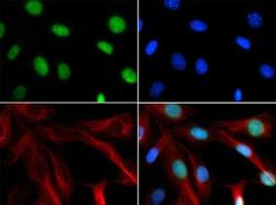
- Immunofluorescent analysis of NIH/3T3 cells using CHD7 polyclonal antibody (Product # PA5-72964) conjugated with FITC (green). Counterstain was performed with FITC (green), DAPI (blue) and Phalloidin (red).
- Submitted by
- Invitrogen Antibodies (provider)
- Main image

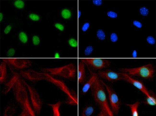
- Experimental details
- Immunofluorescence analysis of CHD7 was performed using 70% confluent log phase SH-SY5Y cells. The cells were fixed with 4% paraformaldehyde for 10 minutes, permeabilized with 0.1% Triton™ X-100 for 15 minutes, and blocked with 2% BSA for 1 hour at room temperature. The cells were labeled with CHD7 Polyclonal Antibody (Product # PA5-72964) at 1:100 dilution in 0.1% BSA, incubated at 4 degree Celsius overnight and then labeled with Goat anti-Rabbit IgG (H+L) Superclonal™ Secondary Antibody, Alexa Fluor® 488 conjugate (Product # A27034) at a dilution of 1:2000 for 45 minutes at room temperature (Panel a: green). Nuclei (Panel b: blue) were stained with ProLong™ Diamond Antifade Mountant with DAPI (Product # P36962). F-actin (Panel c: red) was stained with Rhodamine Phalloidin (Product # R415, 1:300). Panel d represents the merged image showing nuclear localization. Panel e represents cells with no primary antibody to assess background. The images were captured at 60X magnification..
- Submitted by
- Invitrogen Antibodies (provider)
- Main image

- Experimental details
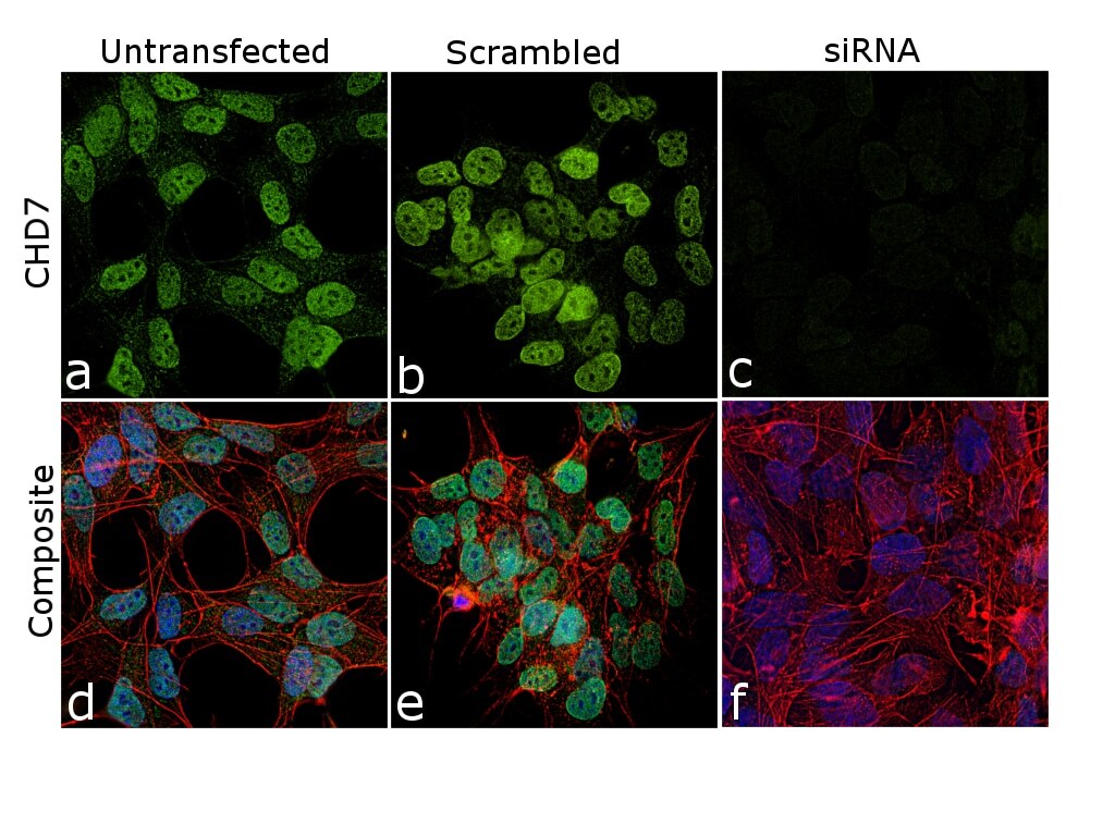
- KD of CHD7 was achieved by transfecting SH-SY5Y cells with CHD7 specific siRNA (Silencer® select Product # s31140, s529331). Immunofluorescence analysis was performed on untransfected SH-SY5Y cells (panel a,d), transfected with non-specific scrambled siRNA (panels b,e) and CHD7 specific siRNA (panel c,f). Cells were fixed, permeabilized, and labelled with CHD7 Polyclonal Antibody (Product # PA5-72964, 1:100 dilution), followed by Goat anti-Rabbit IgG (H+L) Superclonal™ Secondary Antibody, Alexa Fluor® 488 conjugate (Product # A27034, 1:2000). Nuclei (blue) were stained using ProLong™ Diamond Antifade Mountant with DAPI (Product # P36962), and Rhodamine Phalloidin (Product # R415, 1:300) was used for cytoskeletal F-actin (red) staining. Reduction of specific signal was observed upon siRNA mediated KD (panel c,f) confirming specificity of the antibody to CHD7. The images were captured at 60X magnification..
- Submitted by
- Invitrogen Antibodies (provider)
- Main image
- Experimental details
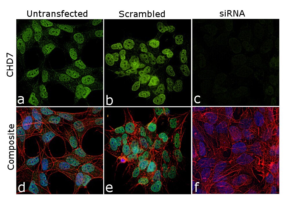
- KD of CHD7 was achieved by transfecting SH-SY5Y cells with CHD7 specific siRNA (Silencer® select Product # s31140, s529331). Immunofluorescence analysis was performed on untransfected SH-SY5Y cells (panel a,d), transfected with non-specific scrambled siRNA (panels b,e) and CHD7 specific siRNA (panel c,f). Cells were fixed, permeabilized, and labelled with CHD7 Polyclonal Antibody (Product # PA5-72964, 1:100 dilution), followed by Goat anti-Rabbit IgG (Heavy Chain) Superclonal™ Secondary Antibody, Alexa Fluor® 488 conjugate (Product # A27034, 1:2000). Nuclei (blue) were stained using ProLong™ Diamond Antifade Mountant with DAPI (Product # P36962), and Rhodamine Phalloidin (Product # R415, 1:300) was used for cytoskeletal F-actin (red) staining. Reduction of specific signal was observed upon siRNA mediated KD (panel c,f) confirming specificity of the antibody to CHD7. The images were captured at 60X magnification..
- Submitted by
- Invitrogen Antibodies (provider)
- Main image

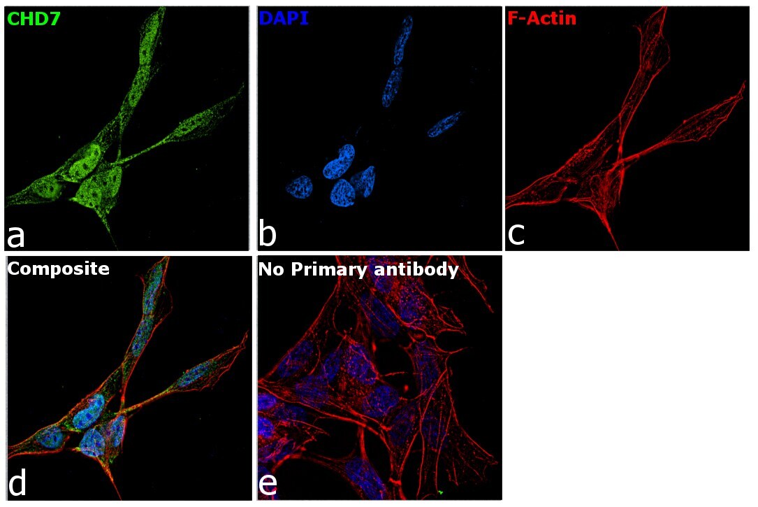
- Experimental details
- Immunofluorescence analysis of CHD7 was performed using 70% confluent log phase SH-SY5Y cells. The cells were fixed with 4% paraformaldehyde for 10 minutes, permeabilized with 0.1% Triton™ X-100 for 15 minutes, and blocked with 2% BSA for 1 hour at room temperature. The cells were labeled with CHD7 Polyclonal Antibody (Product # PA5-72964) at 1:100 dilution in 0.1% BSA, incubated at 4 degree Celsius overnight and then labeled with Goat anti-Rabbit IgG (Heavy Chain) Superclonal™ Secondary Antibody, Alexa Fluor® 488 conjugate (Product # A27034) at a dilution of 1:2000 for 45 minutes at room temperature (Panel a: green). Nuclei (Panel b: blue) were stained with ProLong™ Diamond Antifade Mountant with DAPI (Product # P36962). F-actin (Panel c: red) was stained with Rhodamine Phalloidin (Product # R415, 1:300). Panel d represents the merged image showing nuclear localization. Panel e represents cells with no primary antibody to assess background. The images were captured at 60X magnification..

Supportive validation
- Submitted by
- Invitrogen Antibodies (provider)
- Main image
- Experimental details
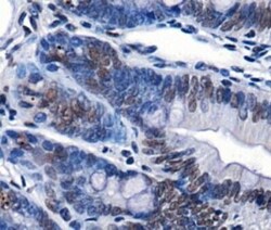
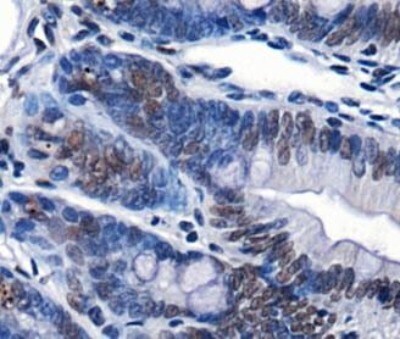
- Immunohistochemical analysis of CHD7 in mouse intestine. Samples were incubated with CHD7 polyclonal antibody (Product # PA5-72964) followed by using DAB with hematoxylin counterstain.
- Submitted by
- Invitrogen Antibodies (provider)
- Main image

- Experimental details
- Immunohistochemical analysis of CHD7 in mouse intestine. Samples were incubated with CHD7 polyclonal antibody (Product # PA5-72964) followed by using DAB with hematoxylin counterstain.

Supportive validation
- Submitted by
- Invitrogen Antibodies (provider)
- Main image
- Experimental details
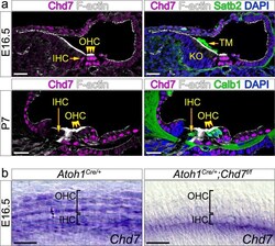
- Fig. 1 Chd7 expression in wildtype and Atoh1 Cre/+ ;Chd7 f/f mutant organ of Corti. a Immunohistochemistry in wildtype cochlea showing Chd7 expression in hair cells at E16.5 and P7. Expression is weaker at P7 compared to E16.5. Hair cells are stained with F-actin and Calbindin 1. Tectorial membrane is stained with Satb2. b In-situ hybridisation showing loss of Chd7 expression in Atoh1 Cre/+ ;Chd7 f/f mutant hair cells within the middle region of the cochlea at E16.5. Atoh1 Cre/+ or Chd7 floxed siblings were used as controls (indicated at the top of the panel). IHC inner hair cells; OHC outer hair cells; TM tectorial membrane. Scale bars 25 um.