Explore
 Validate
 Learn
Learn Western blot
Antibody data
- Antibody Data
- Antigen structure
- References [0]
- Comments [0]
- Validations
- Western blot [1]
- ELISA [1]
- Immunocytochemistry [1]
- Immunohistochemistry [2]
- Flow cytometry [1]
Submit
Validation data
Reference
Comment
Report error
- Product number
- AMM81287 - Provider product page

- Provider
- EnkiLife Biotech Co., Ltd.
- Product name
- PLIN2 Mouse Monoclonal Antibody
- Antibody type
- Monoclonal
- Description
- Affinity Purification
- Reactivity
- Human
- Host
- Mouse
- Conjugate
- Unconjugated
- Antibody clone number
- Monoclonal
- Vial size
- 100 µl
- Concentration
- 1 mg/ml
- Storage
- Store at 4°C short term. Aliquot and store at -20°C long term. Avoid freeze/thaw cycles.
- Handling
- The antibody solution should be gently mixed before use.
No comments: Submit comment
Supportive validation
- Submitted by
- EnkiLife Biotech Co., Ltd. (provider)
- Main image
- Experimental details
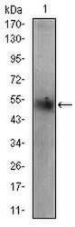
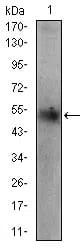
- Western blot analysis using PLIN2 mouse mAb against HepG2 cell lysate.
Supportive validation
- Submitted by
- EnkiLife Biotech Co., Ltd. (provider)
- Main image
- Experimental details
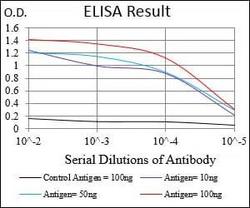
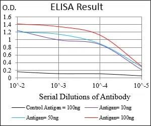
- Black line: Control Antigen (100 ng); Purple line: Antigen(10ng); Blue line: Antigen (50 ng); Red line: Antigen (100 ng);
Supportive validation
- Submitted by
- EnkiLife Biotech Co., Ltd. (provider)
- Main image
- Experimental details
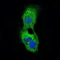
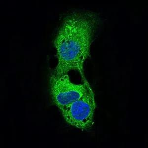
- Immunofluorescence analysis of HepG2 cells using PLIN2 mouse mAb (green). Blue: DRAQ5 fluorescent DNA dye.
Supportive validation
- Submitted by
- EnkiLife Biotech Co., Ltd. (provider)
- Main image
- Experimental details
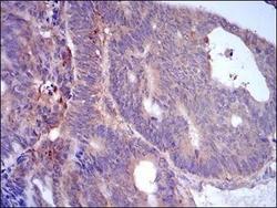
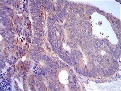
- Immunohistochemical analysis of paraffin-embedded human rectum cancer tissues using PLIN2 mouse mAb with DAB staining.
- Submitted by
- EnkiLife Biotech Co., Ltd. (provider)
- Main image
- Experimental details
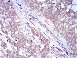
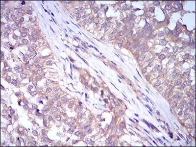
- Immunohistochemical analysis of paraffin-embedded human bladder cancer tissues using PLIN2 mouse mAb with DAB staining.
Supportive validation
- Submitted by
- EnkiLife Biotech Co., Ltd. (provider)
- Main image
- Experimental details
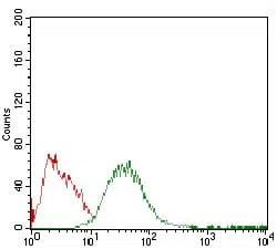
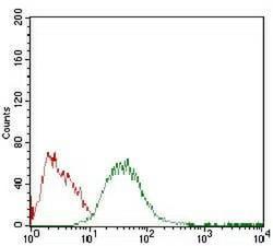
- Flow cytometric analysis of HepG2 cells using PLIN2 mouse mAb (green) and negative control (red).