Explore
 Validate
 Learn
Learn Western blot
 Immunocytochemistry
Antibody data
- Antibody Data
- Antigen structure
- References [2]
- Comments [0]
- Validations
- Immunocytochemistry [3]
- Immunohistochemistry [1]
- Other assay [1]
Submit
Validation data
Reference
Comment
Report error
- Product number
- PA5-34743 - Provider product page

- Provider
- Invitrogen Antibodies
- Product name
- CXCL9 Polyclonal Antibody
- Antibody type
- Polyclonal
- Antigen
- Synthetic peptide
- Description
- Recommended positive controls: A431. Store product as a concentrated solution. Centrifuge briefly prior to opening the vial.
- Reactivity
- Human
- Host
- Rabbit
- Isotype
- IgG
- Vial size
- 100 μL
- Concentration
- 1.33 mg/mL
- Storage
- Store at 4°C short term. For long term storage, store at -20°C, avoiding freeze/thaw cycles.
Submitted references CXCL9 inhibits tumour growth and drives anti-PD-L1 therapy in ovarian cancer.
KCNK3 Mutation Causes Altered Immune Function in Pulmonary Arterial Hypertension Patients and Mouse Models.
Seitz S, Dreyer TF, Stange C, Steiger K, Bräuer R, Scheutz L, Multhoff G, Weichert W, Kiechle M, Magdolen V, Bronger H
British journal of cancer 2022 Jun;126(10):1470-1480
British journal of cancer 2022 Jun;126(10):1470-1480
KCNK3 Mutation Causes Altered Immune Function in Pulmonary Arterial Hypertension Patients and Mouse Models.
West JD, Austin ED, Rizzi EM, Yan L, Tanjore H, Crabtree AL, Moore CS, Muthian G, Carrier EJ, Jacobson DA, Hamid R, Kendall PL, Majka S, Rathinasabapathy A
International journal of molecular sciences 2021 May 9;22(9)
International journal of molecular sciences 2021 May 9;22(9)
No comments: Submit comment
Supportive validation
- Submitted by
- Invitrogen Antibodies (provider)
- Main image
- Experimental details
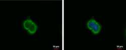
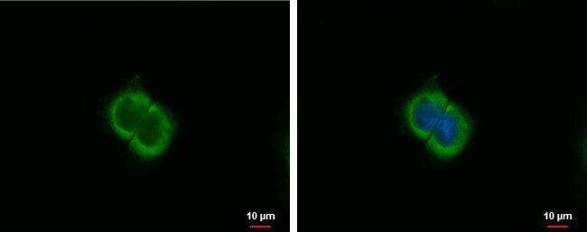
- CXCL9 Polyclonal Antibody detects MIG protein at cytoplasm by immunofluorescent analysis. Sample: A431 cells were fixed in 4% paraformaldehyde at RT for 5 min. Green: MIG protein stained by CXCL9 Polyclonal Antibody (Product # PA5-34743) diluted at 1:1,000. Blue: Hoechst 33342 staining.
- Submitted by
- Invitrogen Antibodies (provider)
- Main image

- Experimental details
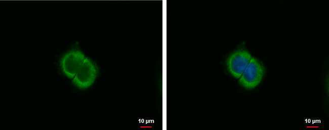
- CXCL9 Polyclonal Antibody detects MIG protein at cytoplasm by immunofluorescent analysis. Sample: A431 cells were fixed in 4% paraformaldehyde at RT for 5 min. Green: MIG protein stained by CXCL9 Polyclonal Antibody (Product # PA5-34743) diluted at 1:1,000. Blue: Hoechst 33342 staining.

- Submitted by
- Invitrogen Antibodies (provider)
- Main image
- Experimental details
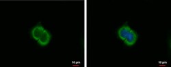
- CXCL9 Polyclonal Antibody detects MIG protein at cytoplasm by immunofluorescent analysis. Sample: A431 cells were fixed in 4% paraformaldehyde at RT for 5 min. Green: MIG protein stained by CXCL9 Polyclonal Antibody (Product # PA5-34743) diluted at 1:1,000. Blue: Hoechst 33342 staining.
Supportive validation
- Submitted by
- Invitrogen Antibodies (provider)
- Main image
- Experimental details
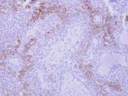
- CXCL9 Polyclonal Antibody detects CXCL9 protein at cytosol on human lung adenocarcinoma by immunohistochemical analysis. Sample: Paraffin-embedded lung adenocarcinoma. CXCL9 Polyclonal Antibody (Product # PA5-34743) dilution: 1:250. Antigen Retrieval: EDTA based buffer, pH 8.0, 15 min.

Supportive validation
- Submitted by
- Invitrogen Antibodies (provider)
- Main image
- Experimental details
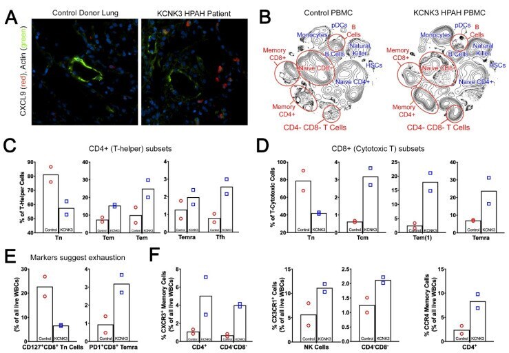
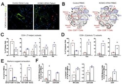
- Figure 6 IHC and CyTOF assessment on human tissue samples validate the role of inflammation in driving KCNK3-PAH. ( A ) Immunofluorescence depicting increased numbers CXCL9 + cells (red) in the KCNK3 patient lung, around the muscularized vessels (green), 400X magnification. ( B ) 2D density contour expression of mass immunotyping of KCNK3 PBMCs. ( C and D ) Naive T cells are downregulated in KCNK3 PBMCs, compensated through the upregulation of central and memory cells in CD4 + and CD8 + population. ( E and F ) CyTOF findings verify the role of cell mediated immune response in KCNK3 subjects through the dysregulation of CXCR3 + , CX3CR1 + , CCR4 + , CD127 + CD8 + and PD1 + CD8 + cells.