Explore
 Validate
 Learn
Learn Western blot
 Immunocytochemistry
Antibody data
- Antibody Data
- Antigen structure
- References [1]
- Comments [0]
- Validations
- Western blot [1]
- Immunohistochemistry [1]
Submit
Validation data
Reference
Comment
Report error
- Product number
- NBP2-12906 - Provider product page

- Provider
- Novus Biologicals
- Product name
- Mouse Monoclonal TRPM7 Antibody
- Antibody type
- Monoclonal
- Description
- Protein G purified. Detects approx 220kDa. No cross-reactivity against TrpM6.
- Reactivity
- Human, Mouse, Rat
- Host
- Mouse
- Isotype
- IgG
- Vial size
- 0.1 mg
- Concentration
- 1 mg/ml
- Storage
- Store at 4C short term. Aliquot and store at -20C long term. Avoid freeze-thaw cycles.
Submitted references TRPM7 is overexpressed in human IBD-related and sporadic colorectal cancer and correlates with tumor grade.
Pugliese D, Armuzzi A, Castri F, Benvenuto R, Mangoni A, Guidi L, Gasbarrini A, Rapaccini GL, Wolf FI, Trapani V
Digestive and liver disease : official journal of the Italian Society of Gastroenterology and the Italian Association for the Study of the Liver 2020 Jun 3;
Digestive and liver disease : official journal of the Italian Society of Gastroenterology and the Italian Association for the Study of the Liver 2020 Jun 3;
No comments: Submit comment
Supportive validation
- Submitted by
- Novus Biologicals (provider)
- Main image
- Experimental details
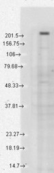
- Western Blot: TRPM7 Antibody (S74-25) [NBP2-12906] - analysis of Human Cell lysates showing detection of TrpM7 protein using Mouse Anti-TrpM7 Monoclonal Antibody, Clone S74-25 . Load: 15 ug protein. Block: 1.5% BSA for 30 minutes at RT. Primary Antibody: Mouse Anti-TrpM7 Monoclonal Antibody at 1:1000 for 2 hours at RT. Secondary Antibody: Sheep Anti-Mouse IgG: HRP for 1 hour at RT.

Supportive validation
- Submitted by
- Novus Biologicals (provider)
- Main image
- Experimental details
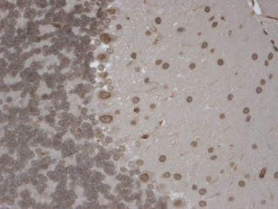
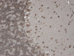
- Immunohistochemistry-Paraffin: TRPM7 Antibody (S74-25) [NBP2-12906] - Tissue: Brain Slice. Species: Mouse. Fixation: 10% Formalin Solution for 12-24 hours at RT. Primary Antibody: Mouse Anti-TrpM7 Monoclonal Antibody at 1:1000 for 1 hour at RT. Secondary Antibody: HRP/DAB Detection System: Biotinylated Goat Anti-Mouse, Streptavidin Peroxidase, DAB Chromogen (brown) for 30 minutes at RT. Counterstain: Mayer Hematoxylin (purple/blue) nuclear stain at 250-500 ul for 5 minutes at RT. Localization: Nuclear staining of both neurons and glia.