 Explore
 Validate
 Learn
Learn Western blot
 Immunocytochemistry
Antibody data
- Antibody Data
- Antigen structure
- References [1]
- Comments [0]
- Validations
- Immunocytochemistry [1]
- Immunohistochemistry [4]
- Other assay [1]
Submit
Validation data
Reference
Comment
Report error
- Product number
- PA5-62621 - Provider product page

- Provider
- Invitrogen Antibodies
- Product name
- GSPT1 Polyclonal Antibody
- Antibody type
- Polyclonal
- Antigen
- Recombinant protein fragment
- Description
- Immunogen sequence: GGAANNHGAG SGAGGRAAPV ESSQEEQSLC EGSNSAVS Highest antigen sequence identity to the following orthologs: Mouse - 66%, Rat - 74%.
- Reactivity
- Human
- Host
- Rabbit
- Isotype
- IgG
- Vial size
- 100 μL
- Concentration
- 0.10 mg/mL
- Storage
- Store at 4°C short term. For long term storage, store at -20°C, avoiding freeze/thaw cycles.
Submitted references Effect of small molecule eRF3 degraders on premature termination codon readthrough.
Baradaran-Heravi A, Balgi AD, Hosseini-Farahabadi S, Choi K, Has C, Roberge M
Nucleic acids research 2021 Apr 19;49(7):3692-3708
Nucleic acids research 2021 Apr 19;49(7):3692-3708
No comments: Submit comment
Supportive validation
- Submitted by
- Invitrogen Antibodies (provider)
- Main image

- Experimental details
- Immunofluorescent staining of GSPT1 in human cell line HeLa shows positivity in cytoplasm & vesicles. Samples were probed using a GSPT1 Polyclonal Antibody (Product # PA5-62621).

Supportive validation
- Submitted by
- Invitrogen Antibodies (provider)
- Main image
- Experimental details
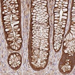
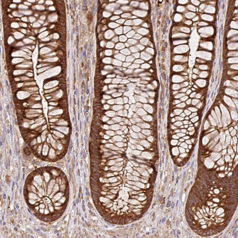
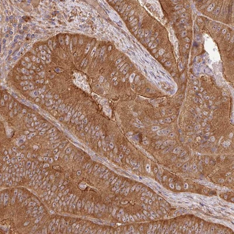
- Immunohistochemical staining of GSPT1 in human rectum using a GSPT1 Polyclonal Antibody (Product # PA5-62621) shows moderate to strong cytoplasmic positivity in glandular cells.
- Submitted by
- Invitrogen Antibodies (provider)
- Main image
- Experimental details
- Immunohistochemical staining of GSPT1 in human testis using a GSPT1 Polyclonal Antibody (Product # PA5-62621) shows moderate to strong cytoplasmic positivity in cells in seminiferous ducts and Leydig cells.

- Submitted by
- Invitrogen Antibodies (provider)
- Main image
- Experimental details
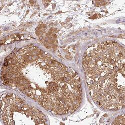
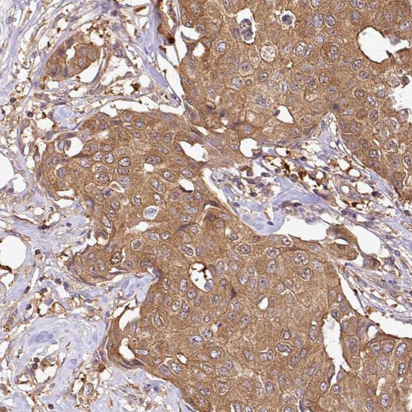
- Immunohistochemical staining of GSPT1 in human breast cancer using a GSPT1 Polyclonal Antibody (Product # PA5-62621) shows moderate to strong cytoplasmic positivity in tumor cells.
- Submitted by
- Invitrogen Antibodies (provider)
- Main image

- Experimental details
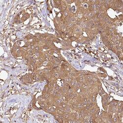
- Immunohistochemical staining of GSPT1 in human colorectal cancer using a GSPT1 Polyclonal Antibody (Product # PA5-62621) shows moderate to strong cytoplasmic positivity in tumor cells.
Supportive validation
- Submitted by
- Invitrogen Antibodies (provider)
- Main image
- Experimental details
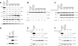
- Figure 1. Effect of siRNA knockdown of eRF3a and eRF3b on TP53 PTC readthrough. ( A ) HDQ-P1 cells with a homozygous nonsense mutation in the TP53 gene (R213X) were transfected with either non-target siRNA or several ON-TARGETplus siRNAs targeting eRF3a . Levels of SURF complex components eRF3a, eRF1, UPF1 and SMG1 were measured 96 h after transfection. GAPDH was used as loading control. ( B ) p53 levels (full-length, FL-p53; truncated, TR-p53) were measured in the same extracts as in A . Vinculin was used as loading control. ( C ) p53 and vinculin levels were measured in transfected cells exposed to 20 mug/ml G418 for 48 h. ( D ) eRF3a and eRF3b levels were measured in HDQ-P1 and H1299-p53 R213X cell extracts. ( E, F ) H1299-p53 R213X cells were transfected with non-target siRNA or several ON-TARGETplus siRNAs targeting eRF3a ( E ) or eRF3b ( F ). eRF3a and eRF3b levels were measured 96 h after transfection. 48 h after transfection, samples were exposed to 20 mug/ml G418 for another 48 h and p53 and vinculin levels were determined. Protein levels in all panels were measured using automated capillary electrophoresis western analysis. In this and subsequent figures, equal amounts of protein lysate were loaded in all capillaries in each panel, except where indicated. The amounts of eRF3a, eRF3b, eRF1, UPF1 and SMG1 are expressed as percentage of levels in untreated cells. As FL-p53 is essentially undetectable in untreated HDQ-P1 cells in most experiments, FL-p53 levels are expr
