Explore
 Validate
 Learn
LearnMA4-001
antibody from Invitrogen Antibodies
Targeting: RASA1
CM-AVM, GAP, p120, p120GAP, p120RASGAP, RASA
 Western blot
 Immunoprecipitation
Antibody data
- Antibody Data
- Antigen structure
- References [3]
- Comments [0]
- Validations
- Western blot [1]
- Immunocytochemistry [6]
Submit
Validation data
Reference
Comment
Report error
- Product number
- MA4-001 - Provider product page

- Provider
- Invitrogen Antibodies
- Product name
- RASA1 Monoclonal Antibody (B4F8)
- Antibody type
- Monoclonal
- Antigen
- Recombinant full-length protein
- Description
- MA4-001 detects GTPase activating protein (GAP) from human, mouse, rat, bovine and non-human primate tissues. MA4-001 has been successfully used in Western blot, immunofluorescence and immunoprecipitation procedures. By Western blot, this antibody recognizes a single 120 kDa protein representing ras GAP from RS-2 cell lysate. Immunofluorescence staining of GAP in mouse fibroblast cells with MA4-001 results in diffuse cytoplasmic staining. Following the addition of PDGF, immunofluorescence staining shows that some GAP rapidly translocates to the plasma membrane. MA4-001 immunoprecipitates GAP that is complexed with the GAP related p190. The MA4-001 antigen is full length recombinant human GAP. Epitope mapping studies suggest that this antibody binds a portion of GAP that contains the src homology regions SH2 & SH3. Reconstitute with PBS.
- Reactivity
- Human, Mouse, Rat, Bovine
- Host
- Mouse
- Isotype
- IgG
- Antibody clone number
- B4F8
- Vial size
- 100 μg
- Concentration
- 1 mg/mL
- Storage
- -20°C, Avoid Freeze/Thaw Cycles
Submitted references Differential expression of cytoskeletal regulatory factors in the adolescent prefrontal cortex: Implications for cortical development.
Molecular cloning of cDNAs encoding the GAP-associated protein p190: implications for a signaling pathway from ras to the nucleus.
Molecular cloning of cDNAs encoding the GAP-associated protein p190: implications for a signaling pathway from ras to the nucleus.
Shapiro LP, Parsons RG, Koleske AJ, Gourley SL
Journal of neuroscience research 2017 May;95(5):1123-1143
Journal of neuroscience research 2017 May;95(5):1123-1143
Molecular cloning of cDNAs encoding the GAP-associated protein p190: implications for a signaling pathway from ras to the nucleus.
Settleman J, Narasimhan V, Foster LC, Weinberg RA
Cell 1992 May 1;69(3):539-49
Cell 1992 May 1;69(3):539-49
Molecular cloning of cDNAs encoding the GAP-associated protein p190: implications for a signaling pathway from ras to the nucleus.
Settleman J, Narasimhan V, Foster LC, Weinberg RA
Cell 1992 May 1;69(3):539-49
Cell 1992 May 1;69(3):539-49
No comments: Submit comment
Supportive validation
- Submitted by
- Invitrogen Antibodies (provider)
- Main image

- Experimental details
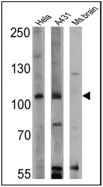
- Western blot analysis of GTPase Activating Protein was performed by loading 25 µg of Hela (Lane 1), A431 (Lane 2), and mouse brain cell lysates (Lane 3) and a molecular weight protein ladder onto an SDS polyacrylamide gel. Proteins were transferred to a PVDF membrane and blocked with a blocking buffer at 4ºC overnight. The membrane was probed with a GTPase Activating Protein monoclonal antibody (Product # MA4-001) at a dilution of 1:200 overnight at 4°C, washed in TBST, and probed with an HRP-conjugated secondary antibody for 1 hr at room temperature in the dark. Chemiluminescent detection was performed using Pierce ECL Plus Western Blotting Substrate (Product # 32132). Results show a band at 110 kDa in all three cell lines.
Supportive validation
- Submitted by
- Invitrogen Antibodies (provider)
- Main image

- Experimental details
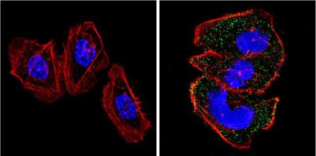
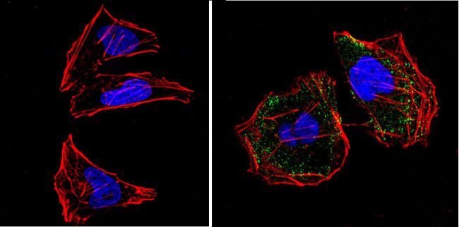
- Immunofluorescent analysis of GTPase Activating Protein (green) showing positive staining in the cytoplasm of C2C12 cells (right) compared with a negative control in the absence of primary antibody (left). Formalin-fixed cells were permeabilized with 0.1% Triton X-100 in TBS for 5-10 minutes, blocked with 3% BSA-PBS for 30 minutes at room temperature and probed with a GTPase Activating Protein monoclonal antibody (Product # MA4-001) in 3% BSA-PBS at a dilution of 1:200 and incubated overnight at 4 ºC in a humidified chamber. Cells were washed with PBST and incubated with a DyLight 488-conjugated goat-anti-mouse IgG (H+L) secondary antibody in PBS at room temperature in the dark. F-actin (red) was stained with a fluorescent red phalloidin and nuclei (blue) were stained with DAPI for 5-10 minutes in the dark. Images were taken at a magnification of 60x.
- Submitted by
- Invitrogen Antibodies (provider)
- Main image

- Experimental details
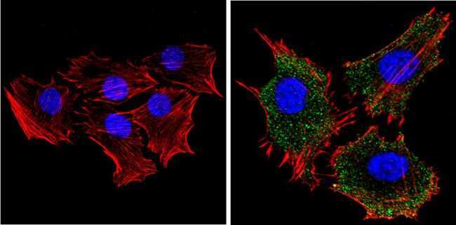
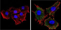
- Immunofluorescent analysis of GTPase Activating Protein (green) showing positive staining in the cytoplasm of Hela cells (right) compared with a negative control in the absence of primary antibody (left). Formalin-fixed cells were permeabilized with 0.1% Triton X-100 in TBS for 5-10 minutes, blocked with 3% BSA-PBS for 30 minutes at room temperature and probed with a GTPase Activating Protein monoclonal antibody (Product # MA4-001) in 3% BSA-PBS at a dilution of 1:200 and incubated overnight at 4 ºC in a humidified chamber. Cells were washed with PBST and incubated with a DyLight 488-conjugated goat-anti-mouse IgG (H+L) secondary antibody in PBS at room temperature in the dark. F-actin (red) was stained with a fluorescent red phalloidin and nuclei (blue) were stained with DAPI for 5-10 minutes in the dark. Images were taken at a magnification of 60x.

- Submitted by
- Invitrogen Antibodies (provider)
- Main image

- Experimental details
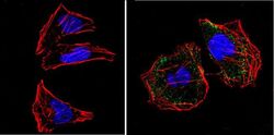
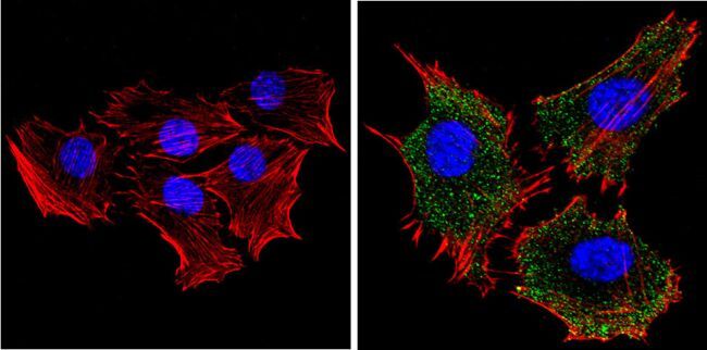
- Immunofluorescent analysis of GTPase Activating Protein (green) showing positive staining in the cytoplasm of A431 cells (right) compared with a negative control in the absence of primary antibody (left). Formalin-fixed cells were permeabilized with 0.1% Triton X-100 in TBS for 5-10 minutes, blocked with 3% BSA-PBS for 30 minutes at room temperature and probed with a GTPase Activating Protein monoclonal antibody (Product # MA4-001) in 3% BSA-PBS at a dilution of 1:200 and incubated overnight at 4 ºC in a humidified chamber. Cells were washed with PBST and incubated with a DyLight 488-conjugated goat-anti-mouse IgG (H+L) secondary antibody in PBS at room temperature in the dark. F-actin (red) was stained with a fluorescent red phalloidin and nuclei (blue) were stained with DAPI for 5-10 minutes in the dark. Images were taken at a magnification of 60x.

- Submitted by
- Invitrogen Antibodies (provider)
- Main image
- Experimental details
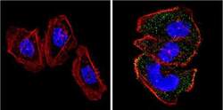
- Immunofluorescent analysis of GTPase Activating Protein (green) showing positive staining in the cytoplasm of Hela cells (right) compared with a negative control in the absence of primary antibody (left). Formalin-fixed cells were permeabilized with 0.1% Triton X-100 in TBS for 5-10 minutes, blocked with 3% BSA-PBS for 30 minutes at room temperature and probed with a GTPase Activating Protein monoclonal antibody (Product # MA4-001) in 3% BSA-PBS at a dilution of 1:200 and incubated overnight at 4 ºC in a humidified chamber. Cells were washed with PBST and incubated with a DyLight 488-conjugated goat-anti-mouse IgG (H+L) secondary antibody in PBS at room temperature in the dark. F-actin (red) was stained with a fluorescent red phalloidin and nuclei (blue) were stained with DAPI for 5-10 minutes in the dark. Images were taken at a magnification of 60x.
- Submitted by
- Invitrogen Antibodies (provider)
- Main image
- Experimental details
- Immunofluorescent analysis of GTPase Activating Protein (green) showing positive staining in the cytoplasm of C2C12 cells (right) compared with a negative control in the absence of primary antibody (left). Formalin-fixed cells were permeabilized with 0.1% Triton X-100 in TBS for 5-10 minutes, blocked with 3% BSA-PBS for 30 minutes at room temperature and probed with a GTPase Activating Protein monoclonal antibody (Product # MA4-001) in 3% BSA-PBS at a dilution of 1:200 and incubated overnight at 4 ºC in a humidified chamber. Cells were washed with PBST and incubated with a DyLight 488-conjugated goat-anti-mouse IgG (H+L) secondary antibody in PBS at room temperature in the dark. F-actin (red) was stained with a fluorescent red phalloidin and nuclei (blue) were stained with DAPI for 5-10 minutes in the dark. Images were taken at a magnification of 60x.
- Submitted by
- Invitrogen Antibodies (provider)
- Main image
- Experimental details
- Immunofluorescent analysis of GTPase Activating Protein (green) showing positive staining in the cytoplasm of A431 cells (right) compared with a negative control in the absence of primary antibody (left). Formalin-fixed cells were permeabilized with 0.1% Triton X-100 in TBS for 5-10 minutes, blocked with 3% BSA-PBS for 30 minutes at room temperature and probed with a GTPase Activating Protein monoclonal antibody (Product # MA4-001) in 3% BSA-PBS at a dilution of 1:200 and incubated overnight at 4 ºC in a humidified chamber. Cells were washed with PBST and incubated with a DyLight 488-conjugated goat-anti-mouse IgG (H+L) secondary antibody in PBS at room temperature in the dark. F-actin (red) was stained with a fluorescent red phalloidin and nuclei (blue) were stained with DAPI for 5-10 minutes in the dark. Images were taken at a magnification of 60x.