Explore
 Validate
 Learn
Learn Western blot
 Chromatin Immunoprecipitation
Antibody data
- Antibody Data
- Antigen structure
- References [0]
- Comments [0]
- Validations
- Western blot [2]
- Immunocytochemistry [1]
- Immunohistochemistry [2]
- Other assay [1]
Submit
Validation data
Reference
Comment
Report error
- Product number
- UM500020 - Provider product page

- Provider
- Invitrogen Antibodies
- Product name
- XPF Monoclonal Antibody (UMAB20), UltraMAB™
- Antibody type
- Monoclonal
- Antigen
- Recombinant full-length protein
- Reactivity
- Human, Mouse, Rat, Canine
- Host
- Mouse
- Isotype
- IgG
- Antibody clone number
- UMAB20
- Vial size
- 100 µL
- Concentration
- 0.5-1.0 mg/mL
- Storage
- -20° C, Avoid Freeze/Thaw Cycles
No comments: Submit comment
Supportive validation
- Submitted by
- Invitrogen Antibodies (provider)
- Main image
- Experimental details
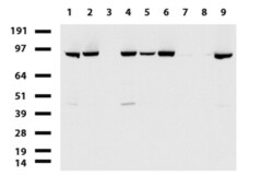
- Western blot of cell lysates (35 µg) from 9 different cell lines (1: HepG2, 2: HeLa, 3: SV-T2, 4: A549, 5: COS7, 6: Jurkat, 7: MDCK, 8: PC-12, 9: MCF7). Dilution: 1:500.

- Submitted by
- Invitrogen Antibodies (provider)
- Main image
- Experimental details
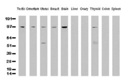
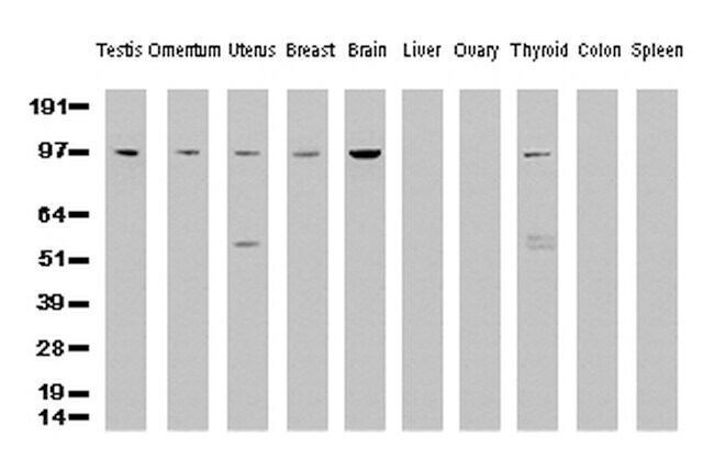
- Western Blot analysis of 10 different human tissue lysates (10 µg) by using anti-XPF monoclonal antibody (clone UMAB20, 1:500)
Supportive validation
- Submitted by
- Invitrogen Antibodies (provider)
- Main image
- Experimental details
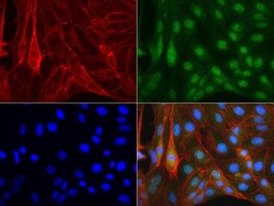
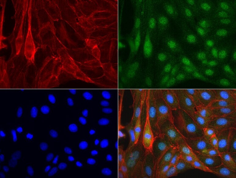
- Immunofluorescent staining of MDCK cells using anti-XPF mouse monoclonal antibody (UM500020, green, 1:50). Actin filaments were labeled with Alexa Fluor 594 Phalloidin (red), and nuclear with DAPI (blue).
Supportive validation
- Submitted by
- Invitrogen Antibodies (provider)
- Main image
- Experimental details
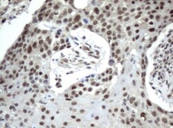
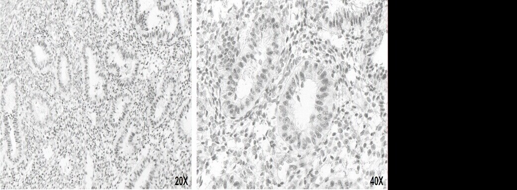
- Immunohistochemical staining of paraffin-embedded Carcinoma of lung tissue using anti-XPF (UMAB20) mouse monoclonal antibody. (UM500020, Dilution 1:50; heat-induced epitope retrieval by 10mM citric buffer, pH6.0, 120°C for 3min)
- Submitted by
- Invitrogen Antibodies (provider)
- Main image
- Experimental details
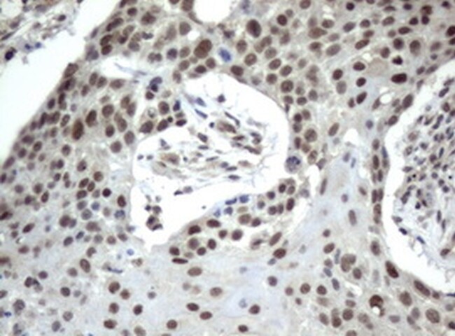
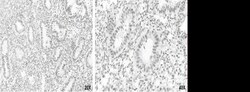
- Immunohistochemical staining of paraffin-embedded human uterus using anti-XPF clone UMAB20 mouse monoclonal antibody (UM500020) 1:50 with Polink2 Broad HRP DAB detection kit; heat-induced epitope retrieval with TEE pH9.0 HIER buffer using pressure chamber for 3 minutes at 110°C. Nuclear staining is seen in the epithiel cells of uterine gland
Supportive validation
- Submitted by
- Invitrogen Antibodies (provider)
- Main image
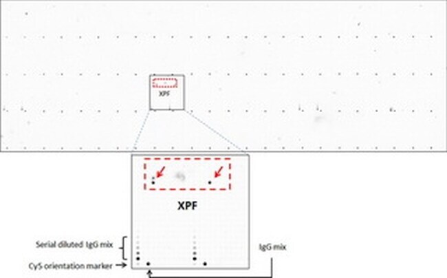
- Experimental details
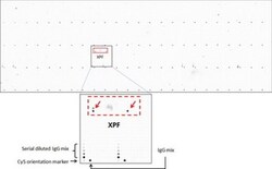
- OriGene overexpression protein microarray chip was immunostained with UltraMAB anti-XPF mouse monoclonal antibody (clone UMAB20). The positive reactive proteins are highlighted with two red arrows in the enlarged subarray. All the positive controls spotted in this subarray are also labeled for clarification. These data show that UltraMAB anti-XPF (UMAB20) very specifically recognizes XPF antigen on OriGene protein microarray chip.