Explore
 Validate
 Learn
Learn Western blot
Antibody data
- Antibody Data
- Antigen structure
- References [1]
- Comments [0]
- Validations
- Western blot [3]
- Immunohistochemistry [1]
- Other assay [1]
Submit
Validation data
Reference
Comment
Report error
- Product number
- PA5-38789 - Provider product page

- Provider
- Invitrogen Antibodies
- Product name
- Anti-XRN2 Polyclonal Antibody
- Antibody type
- Polyclonal
- Antigen
- Synthetic peptide
- Reactivity
- Human, Mouse
- Host
- Rabbit
- Isotype
- IgG
- Vial size
- 100 µg
- Concentration
- 1 mg/mL
- Storage
- -20°C
Submitted references Elevated Wall Tension Leads to Reduced miR-133a in the Thoracic Aorta by Exosome Release.
Akerman AW, Blanding WM, Stroud RE, Nadeau EK, Mukherjee R, Ruddy JM, Zile MR, Ikonomidis JS, Jones JA
Journal of the American Heart Association 2019 Jan 8;8(1):e010332
Journal of the American Heart Association 2019 Jan 8;8(1):e010332
No comments: Submit comment
Supportive validation
- Submitted by
- Invitrogen Antibodies (provider)
- Main image
- Experimental details
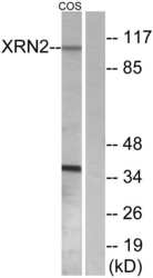
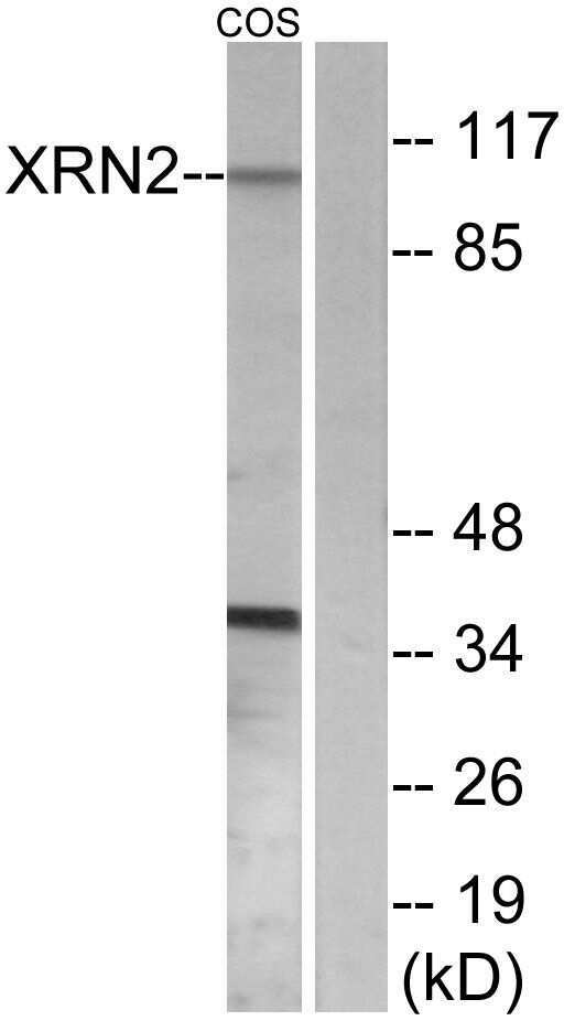
- Western blot analysis of XRN2 in extracts from COS7 cells using a XRN2 polyclonal antibody (Product # PA5-38789).
- Submitted by
- Invitrogen Antibodies (provider)
- Main image

- Experimental details
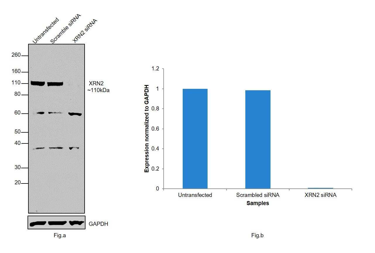
- Knockdown of XRN2 was achieved by transfecting HeLa with XRN2 specific siRNAs (Silencer® select Products # s22413, s22412). Western blot analysis (Fig. a) was performed using whole cell extracts from the XRN2 knockdown cells (Lane 3), non-specific scrambled siRNA transfected cells (Lane 2) and untransfected cells (Lane 1). The blot was probed with XRN2 Polyclonal Antibody (Product # PA5-38789, 1:1000 dilution) and Goat anti-Rabbit IgG (H+L) Superclonal™ Secondary Antibody, HRP conjugate (Product # A27036, 0.25 µg/mL, 1:4000 dilution). Densitometric analysis of this western blot is shown in histogram (Fig. b). Decrease in signal upon siRNA mediated knock down confirms that antibody is specific to XRN2.
- Submitted by
- Invitrogen Antibodies (provider)
- Main image
- Experimental details
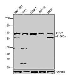
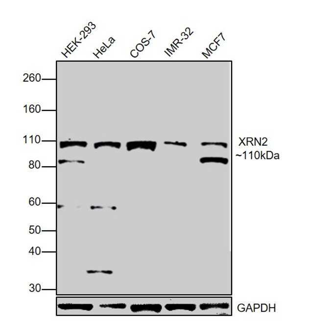
- Western blot was performed using Anti-XRN2 Polyclonal Antibody (Product # PA5-38789) and a 110kDa band corresponding to XRN2 was observed in all the tested cell models. Modified whole cell lysates (1% SDS) (30ug lysate) of HEK-293 (Lane 1), HeLa (Lane 2), COS-7 (Lane 3), IMR-32 (Lane 4) and MCF7 (Lane 5) were electrophoresed using Novex® NuPAGE® 10% Bis-Tris gel (Product # NP0302BOX). Resolved proteins were then transferred onto a nitrocellulose membrane (Product # IB23001) by iBlot® 2 Dry Blotting System (Product # IB21001). The blot was probed with the primary antibody (1:1000 dilution) and detected by chemiluminescence with Goat Anti-Rabbit IgG Secondary Antibody, HRP conjugate (Product # A27036, 1:4000 dilution) using the iBright FL 1000 (Product # A32752). Chemiluminescent detection was performed using SuperSignal™ West Dura Extended Duration Substrate (Product # 34076).
Supportive validation
- Submitted by
- Invitrogen Antibodies (provider)
- Main image
- Experimental details
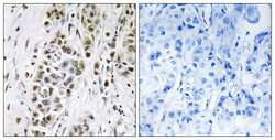
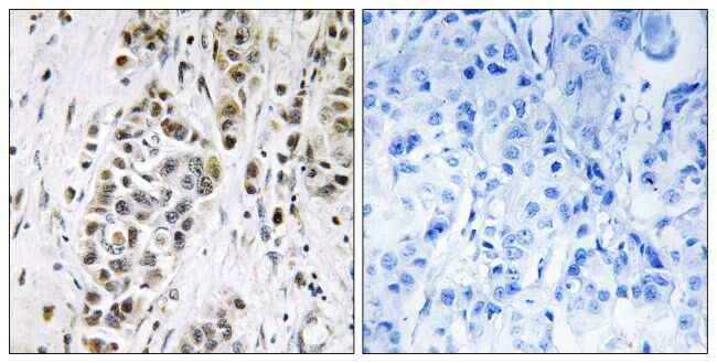
- Immunohistochemical analysis of XRN2 in paraffin-embedded human breast carcinoma using a XRN2 polyclonal antibody (Product # PA5-38789).
Supportive validation
- Submitted by
- Invitrogen Antibodies (provider)
- Main image
- Experimental details
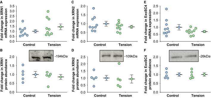
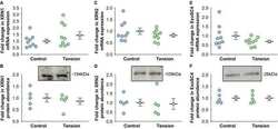
- Figure 4 Mechanical tension and micro RNA- specific ribonuclease mRNA expression and protein abundance. Total RNA or protein was isolated from each independent cell line and examined by real-time polymerase chain reaction or Western blotting, respectively. A , XRN 1 mRNA expression levels in aortic fibroblasts exposed to 12% biaxial cyclic stretch (Tension, n=9) or held static (Control, n=9) for 3 hours. B , XRN 1 protein abundance and representative immunoblot in aortic fibroblasts exposed to 12% biaxial cyclic stretch (Tension, n=5) or held static (Control, n=5) for 3 hours. C , XRN 2 mRNA expression levels in aortic fibroblasts exposed to 12% biaxial cyclic stretch (Tension, n=9) or held static (Control, n=9) for 3 hours. D , XRN 2 protein abundance and representative immunoblot in aortic fibroblasts exposed to 12% biaxial cyclic stretch (Tension, n=5) or held static (Control, n=5) for 3 hours. E , Exo SC 4 mRNA expression levels in aortic fibroblasts exposed to 12% biaxial cyclic stretch (Tension, n=9) or held static (Control, n=9) for 3 hours. F , Exo SC 4 protein abundance and representative immunoblot in aortic fibroblasts exposed to 12% biaxial cyclic stretch (Tension, n=5) or held static (Control, n=5) for 3 hours. A through F , Data are represented in dot plots with the mean and SEM shown next to each group. Comparisons were made using a 2-sample t test (unpaired, 2 tailed). No significant differences were observed vs control.