Explore
 Validate
 Learn
Learn Western blot
Antibody data
- Antibody Data
- Antigen structure
- References [1]
- Comments [0]
- Validations
- Western blot [4]
- Immunoprecipitation [1]
- Immunohistochemistry [3]
Submit
Validation data
Reference
Comment
Report error
- Product number
- NB100-57541 - Provider product page

- Provider
- Novus Biologicals
- Proper citation
- Novus Cat#NB100-57541, RRID:AB_2288770
- Product name
- Rabbit Polyclonal XRN2 Antibody
- Antibody type
- Polyclonal
- Description
- Immunogen affinity purified.
- Reactivity
- Human, Mouse
- Host
- Rabbit
- Isotype
- IgG
- Vial size
- 0.1 ml
- Concentration
- 0.2 mg/ml
- Storage
- Store at 4C. Do not freeze.
Submitted references Changes in cellular mRNA stability, splicing, and polyadenylation through HuR protein sequestration by a cytoplasmic RNA virus.
Barnhart MD, Moon SL, Emch AW, Wilusz CJ, Wilusz J
Cell reports 2013 Nov 27;5(4):909-17
Cell reports 2013 Nov 27;5(4):909-17
No comments: Submit comment
Supportive validation
- Submitted by
- Novus Biologicals (provider)
- Main image
- Experimental details
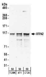
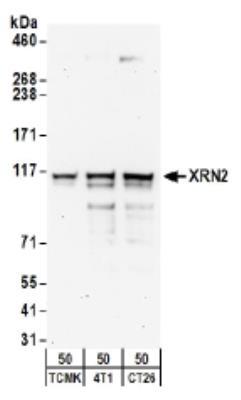
- Western Blot: XRN2 Antibody [NB100-57541] - Detection of Mouse : Whole cell lysate (50 ug) from TCMK-1, 4T1, and CT26.WT cells. Antibodies: Affinity purified rabbit anti-XRN2 antibody used for WB at 0.4 ug/ml.Detection: Chemiluminescence with an exposure time of 3 minutes.
- Submitted by
- Novus Biologicals (provider)
- Main image

- Experimental details
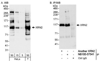
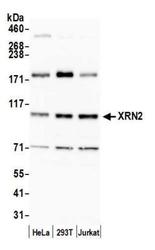
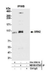
- Western Blot: XRN2 Antibody [NB100-57541] - Whole cell lysate from HeLa (5, 15 and 50 mcg for WB; 1 mg for IP, 20% of IP loaded) and 293T (T; 50 mcg) cells.Affinity purified rabbit anti-XRN2 antibody used for WB at 0.04 mcg/ml (A) and 1 mcg/ml (B) and used for IP at 3 mcg/mg lysate. XRN2 was also immunoprecipitated by rabbit anti-XRN2 antibody, which recognizes an upstream epitope. Detection: Chemiluminescence with exposure times of 3 seconds (A) and 10 seconds (B).
- Submitted by
- Novus Biologicals (provider)
- Main image
- Experimental details
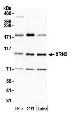
- Western Blot: XRN2 Antibody [NB100-57541] - Detection of human XRN2 by western blot. Samples: Whole cell lysate (15 ug) from HeLa, HEK293T, and Jurkat cells prepared using NETN lysis buffer. Antibody: Affinity purified rabbit anti-XRN2 antibody NB100-57541 used for WB at 0.04 ug/ml. Detection: Chemiluminescence with an exposure time of 30 seconds.
- Submitted by
- Novus Biologicals (provider)
- Main image
- Experimental details
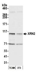
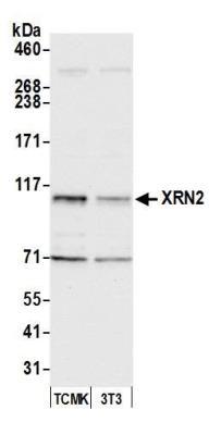
- Western Blot: XRN2 Antibody [NB100-57541] - Detection of mouse XRN2 by western blot. Samples: Whole cell lysate (15 ug) from TCMK-1 and NIH 3T3 cells prepared using NETN lysis buffer. Antibody: Affinity purified rabbit anti-XRN2 antibody NB100-57541 used for WB at 0.1 ug/ml. Detection: Chemiluminescence with an exposure time of 10 seconds.
Supportive validation
- Submitted by
- Novus Biologicals (provider)
- Main image
- Experimental details
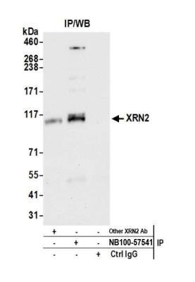
- Immunoprecipitation: XRN2 Antibody [NB100-57541] - Detection of human XRN2 by western blot of immunoprecipitates. Samples: Whole cell lysate (1.0 mg per IP reaction; 20% of IP loaded) from HeLa cells prepared using NETN lysis buffer. Antibodies: Affinity purified rabbit anti-XRN2 antibody NB100-57541 used for IP at 3 ug per reaction. XRN2 was also immunoprecipitated by another rabbit anti-XRN2 antibody. For blotting immunoprecipitated XRN2, NB100-57541 was used at 1 ug/ml. Detection: Chemiluminescence with an exposure time of 10 seconds.
Supportive validation
- Submitted by
- Novus Biologicals (provider)
- Main image
- Experimental details
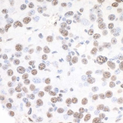
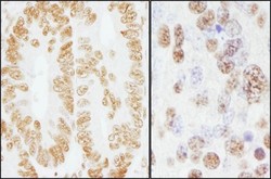
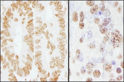
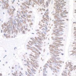
- Immunohistochemistry: XRN2 Antibody [NB100-57541] - Sample: FFPE sections of human colon carcinoma (left) and mouse teratoma (right). Antibody: Affinity purified rabbit anti- XRN2 used at a dilution of 1:200 (1ug/ml). Detection: DAB
- Submitted by
- Novus Biologicals (provider)
- Main image
- Experimental details
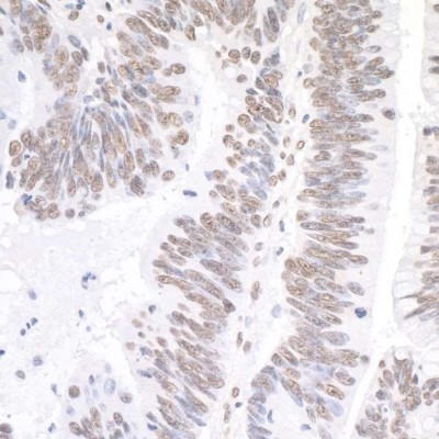
- Immunohistochemistry-Paraffin: XRN2 Antibody [NB100-57541] - Sample: FFPE sections of human colon carcinoma. Antibody: Affinity purified rabbit anti-XRN2 (NB100-57541). Detection: DAB
- Submitted by
- Novus Biologicals (provider)
- Main image
- Experimental details
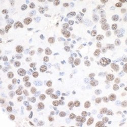
- Immunohistochemistry-Paraffin: XRN2 Antibody [NB100-57541] - Sample: FFPE sections of mouse renal cell carcinoma. Antibody: Affinity purified rabbit anti-XRN2 (NB100-57541). Detection: DAB