Explore
 Validate
 Learn
Learn Western blot
Antibody data
- Antibody Data
- Antigen structure
- References [1]
- Comments [0]
- Validations
- Western blot [3]
- Immunohistochemistry [1]
- Other assay [1]
Submit
Validation data
Reference
Comment
Report error
- Product number
- PA5-45721 - Provider product page

- Provider
- Invitrogen Antibodies
- Product name
- RPS28 Polyclonal Antibody
- Antibody type
- Polyclonal
- Antigen
- Synthetic peptide
- Description
- Peptide sequence: RTGSQGQCTQ VRVEFMDDTS RSIIRNVKGP VREGDVLTLL ESEREARRLR Sequence homology: Cow: 100%; Goat: 100%; Guinea Pig: 100%; Horse: 100%; Human: 100%; Mouse: 100%; Rabbit: 100%; Rat: 100%; Yeast: 93%; Zebrafish: 100%
- Reactivity
- Human
- Host
- Rabbit
- Isotype
- IgG
- Vial size
- 100 μL
- Concentration
- 0.5 mg/mL
- Storage
- -20°C, Avoid Freeze/Thaw Cycles
Submitted references Proteomic and mechanistic dissection of the poxvirus-customized ribosome.
DiGiuseppe S, Rollins MG, Astar H, Khalatyan N, Savas JN, Walsh D
Journal of cell science 2020 Jul 9;134(5)
Journal of cell science 2020 Jul 9;134(5)
No comments: Submit comment
Supportive validation
- Submitted by
- Invitrogen Antibodies (provider)
- Main image
- Experimental details
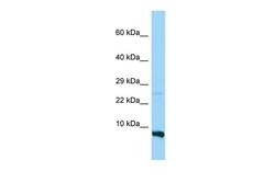
- Western blot analysis of human MDA-MB435 cells using an anti-RPS28 polyclonal antibody (Product # PA5-45721).

- Submitted by
- Invitrogen Antibodies (provider)
- Main image
- Experimental details
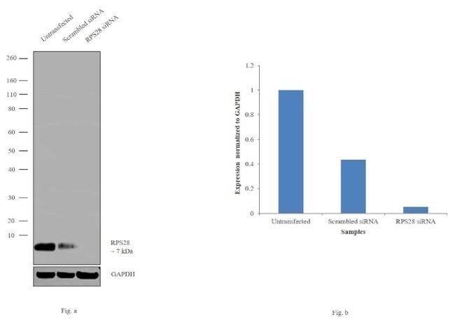
- Knockdown of RPS28 was achieved by transfecting HeLa cells with RPS28 specific siRNAs (Silencer® select Product # s226977). Western blot analysis (Fig. a) was performed using whole cell extracts from the RPS28 knockdown cells (lane 3), non-specific scrambled siRNA transfected cells (lane 2) and untransfected cells (lane 1). The blot was probed with RPS28 Polyclonal Antibody (Product # PA5-45721, 1 µg/mL) and Goat anti-Rabbit IgG (Heavy Chain) Superclonal™ Secondary Antibody, HRP conjugate (Product # A27036, 0.25 µg/mL, 1:4,000 dilution). Densitometric analysis of this western blot is shown in histogram (Fig. b). Decrease in signal upon siRNA mediated knock down confirms that antibody is specific to RPS28.
- Submitted by
- Invitrogen Antibodies (provider)
- Main image
- Experimental details
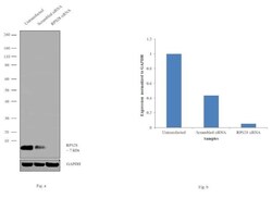
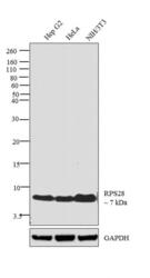
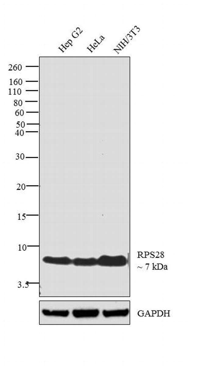
- Western blot analysis was performed on whole cell extracts (30 µg lysate) of Hep G2 (Lane 1), HeLa (Lane 2) and NIH/3T3 (Lane 3). The blot was probed with Anti-RPS28 Polyclonal Antibody (Product # PA5-45721, 1 µg/mL) and detected by chemiluminescence using Goat anti-Rabbit IgG (Heavy Chain) Superclonal™ Secondary Antibody, HRP conjugate (Product # A27036, 0.25 µg/mL, 1:4,000 dilution). A 7 kDa band corresponding to RPS28 was observed across the cell lines tested.
Supportive validation
- Submitted by
- Invitrogen Antibodies (provider)
- Main image
- Experimental details
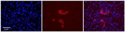
- Immunohistochemistry (paraffin-embedded) analysis of human adult liver tissue using an anti-RPS28 polyclonal antibody (Product # PA5-45721).

Supportive validation
- Submitted by
- Invitrogen Antibodies (provider)
- Main image
- Experimental details
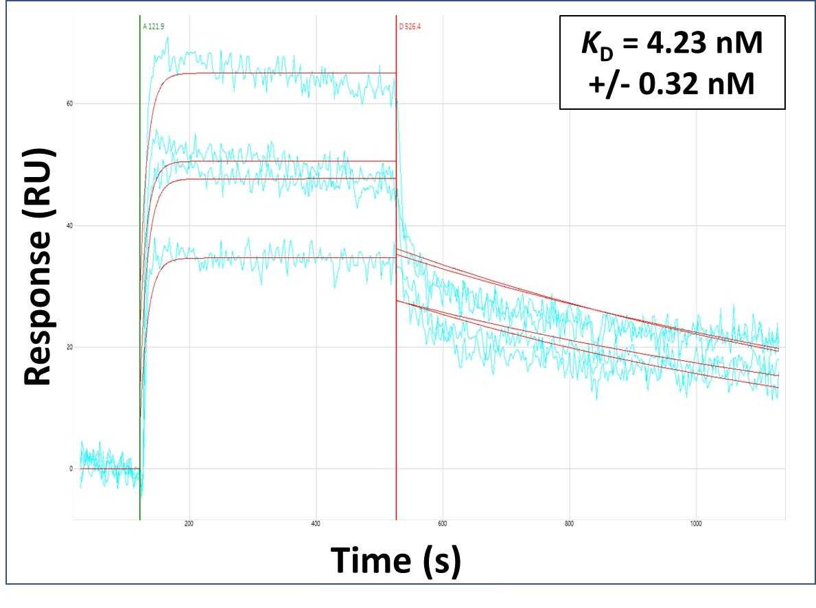
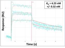
- Surface Plasmon Resonance of RPS28 polyclonal antibody (Product # PA5-45721). Purified polyclonal antibodies were immobilized on a Protein A/G coated Carterra LSA sensor chip at concentrations of 5, and 50 µg/mL in duplicate. Antibodies on the surface were exposed to interaction with peptides sequentially via microfluidic controlled flow at 333 nm peptide concentration for kinetic characterization of the binders for affinity and specificity, followed by curve fitting using the Kinetics software. Kd determinations for both concentrations were averaged and results and standard deviation are shown.