 Explore
 Validate
 Learn
Learn Western blot
Antibody data
- Antibody Data
- Antigen structure
- References [2]
- Comments [0]
- Validations
- Western blot [7]
- Immunocytochemistry [6]
- Immunohistochemistry [12]
- Other assay [6]
Submit
Validation data
Reference
Comment
Report error
- Product number
- PA5-31348 - Provider product page

- Provider
- Invitrogen Antibodies
- Product name
- RAB11B Polyclonal Antibody
- Antibody type
- Polyclonal
- Antigen
- Recombinant full-length protein
- Description
- Recommended positive controls: U87-MG, SK-N-SH, IMR32, SK-N-AS, Mouse brain, PC-12, Rat2. Predicted reactivity: Mouse (100%), Rat (100%), Pig (100%), Chicken (99%), Rhesus Monkey (100%), Bovine (100%). Store product as a concentrated solution. Centrifuge briefly prior to opening the vial.
- Reactivity
- Human, Mouse, Rat
- Host
- Rabbit
- Isotype
- IgG
- Vial size
- 100 μL
- Concentration
- 1.67 mg/mL
- Storage
- Store at 4°C short term. For long term storage, store at -20°C, avoiding freeze/thaw cycles.
Submitted references Rab11b-mediated integrin recycling promotes brain metastatic adaptation and outgrowth.
A novel liver metastasis-correlated protein of pancreatic neuroendocrine neoplasm (PanNEN) discovered by proteomic analysis.
Howe EN, Burnette MD, Justice ME, Schnepp PM, Hedrick V, Clancy JW, Guldner IH, Lamere AT, Li J, Aryal UK, D'Souza-Schorey C, Zartman JJ, Zhang S
Nature communications 2020 Jun 15;11(1):3017
Nature communications 2020 Jun 15;11(1):3017
A novel liver metastasis-correlated protein of pancreatic neuroendocrine neoplasm (PanNEN) discovered by proteomic analysis.
Shimura M, Mizuma M, Takadate T, Katoh Y, Suzuki T, Iseki M, Hata T, Aoki S, Suzuki Y, Sakata N, Ohtsuka H, Hayashi H, Morikawa T, Nakagawa K, Motoi F, Naitoh T, Igarashi K, Sasano H, Unno M
Oncotarget 2018 May 11;9(36):24291-24303
Oncotarget 2018 May 11;9(36):24291-24303
No comments: Submit comment
Supportive validation
- Submitted by
- Invitrogen Antibodies (provider)
- Main image
- Experimental details
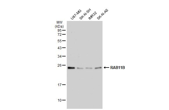
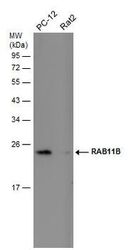
- Western Blot using RAB11B Polyclonal Antibody (Product # PA5-31348). Various whole cell extracts (30 µg) were separated by 12% SDS-PAGE, and the membrane was blotted with RAB11B Polyclonal Antibody (Product # PA5-31348) diluted at 1:1,000. The HRP-conjugated anti-rabbit IgG antibody was used to detect the primary antibody.
- Submitted by
- Invitrogen Antibodies (provider)
- Main image
- Experimental details
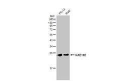
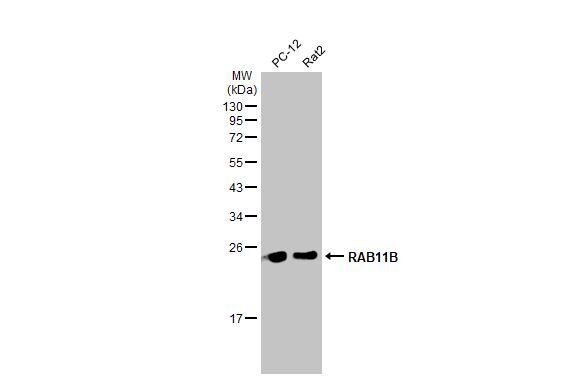
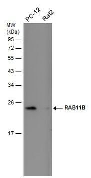
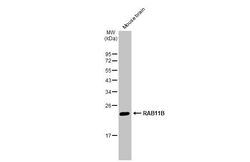
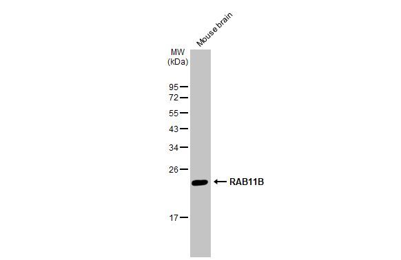
- Western Blot using RAB11B Polyclonal Antibody (Product # PA5-31348). Mouse tissue extract (50 µg) was separated by 12% SDS-PAGE, and the membrane was blotted with RAB11B Polyclonal Antibody (Product # PA5-31348) diluted at 1:1,000. The HRP-conjugated anti-rabbit IgG antibody was used to detect the primary antibody.
- Submitted by
- Invitrogen Antibodies (provider)
- Main image
- Experimental details
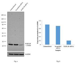
- Knockdown of RAB11B was achieved by transfecting HeLa cells with RAB11B specific siRNAs (Silencer® select Product # s17649). Western blot analysis (Fig. a) was performed using membrane enriched cell extracts from the RAb11B knockdown cells (lane 3), non-specific scrambled siRNA transfected cells (lane 2) and untransfected cells (lane 1). The blots were probed with RAB11B Polyclonal Antibody (Product # PA5-31348, 1:2,000 dilution) Goat anti-Rabbit IgG (Heavy Chain) Superclonal™ Secondary Antibody, HRP conjugate (Product # A27036, 0.25 µg/mL, 1:4,000 dilution). Densitometric analysis of this western blot is shown in histogram (Fig. b). Decrease in signal upon siRNA mediated knock down confirms that antibody is specific to RAB11B.

- Submitted by
- Invitrogen Antibodies (provider)
- Main image
- Experimental details
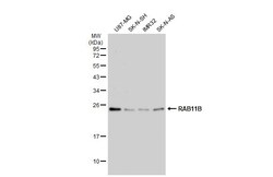
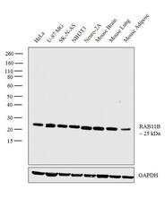
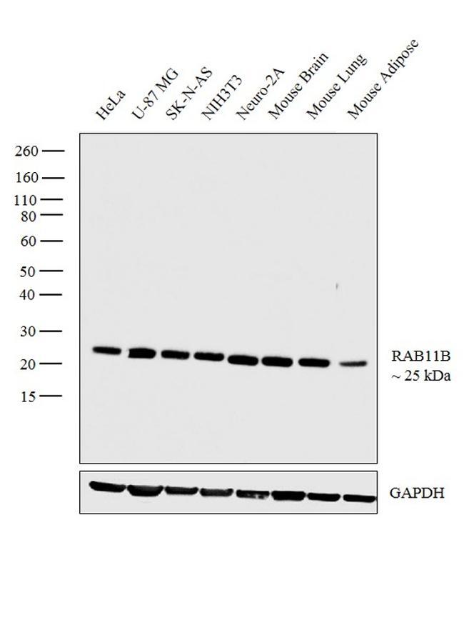
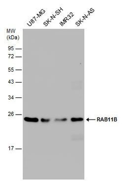
- Western blot analysis was performed on membrane cell extracts (30 µg lysate) of HeLa (Lane 1), U-87 MG (Lane 2), SK-N-AS (Lane 3), NIH3T3 (Lane 4), Neuro-2A (Lane 5), tissue extracts of Mouse Brain (Lane 6), Mouse Lung (Lane 7) and Mouse Adipose (Lane 8). The blot was probed with Anti-RAB11B Polyclonal Antibody (Product # PA5-31348, 1:2,000 dilution) and detected by chemiluminescence using Goat anti-Rabbit IgG (Heavy Chain) Superclonal™ Secondary Antibody, HRP conjugate (Product # A27036, 0.25 µg/mL, 1:4,000 dilution). A 25 kDa band corresponding to RAB11B was observed across all the cell lines and mouse tissue extracts tested.
- Submitted by
- Invitrogen Antibodies (provider)
- Main image
- Experimental details
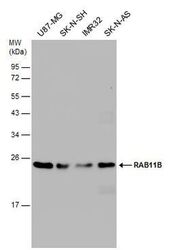
- Western Blot analysis of RAB11B was performed by separating 30 µg of various whole cell extracts by 12% SDS-PAGE. Proteins were transferred to a membrane and probed with a RAB11B Polyclonal Antibody (Product # PA5-31348) at a dilution of 1:1000 and a HRP-conjugated anti-rabbit IgG secondary antibody.
- Submitted by
- Invitrogen Antibodies (provider)
- Main image
- Experimental details
- Western Blot analysis of RAB11B was performed by separating 30 µg of various whole cell extracts by 12% SDS-PAGE. Proteins were transferred to a membrane and probed with a RAB11B Polyclonal Antibody (Product # PA5-31348) at a dilution of 1:1000.
Supportive validation
- Submitted by
- Invitrogen Antibodies (provider)
- Main image
- Experimental details
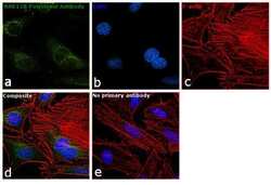
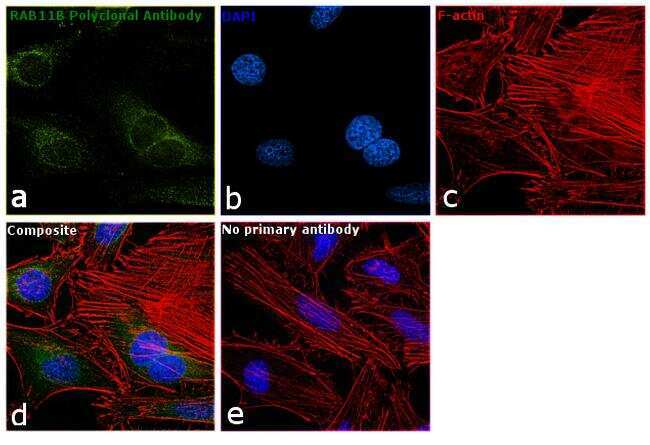
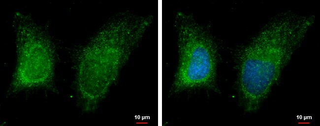
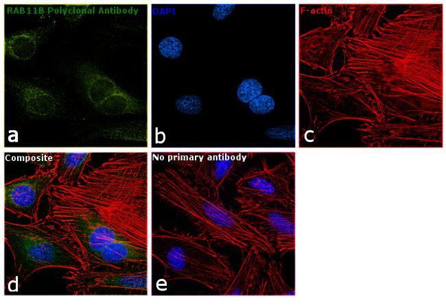
- Immunofluorescence analysis of RAB11B was performed using 70% confluent log phase HeLa cells. The cells were fixed with 4% paraformaldehyde for 10 minutes, permeabilized with 0.1% Triton™ X-100 for 15 minutes, and blocked with 1% BSA for 1 hour at room temperature. The cells were labeled with RAB11B Polyclonal Antibody (Product # PA5-31348) at 1:500 dilution in 0.1% BSA, incubated at 4 degree Celsius overnight and then labeled with Goat anti-Rabbit IgG (H+L) Superclonal™ Secondary Antibody, Alexa Fluor® 488 conjugate (Product # A27034) at a dilution of 1:2000 for 45 minutes at room temperature (Panel a: green). Nuclei (Panel b: blue) were stained with SlowFade® Gold Antifade Mountant with DAPI (Product # S36938). F-actin (Panel c: red) was stained with Rhodamine Phalloidin (Product # R415, 1:300). Panel d represents the merged image showing cytoplasmic localization. Panel e represents control cells with no primary antibody to assess background. The images were captured at 60X magnification.
- Submitted by
- Invitrogen Antibodies (provider)
- Main image

- Experimental details
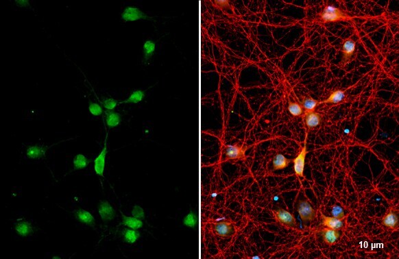
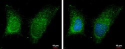
- RAB11B Polyclonal Antibody detects RAB11B protein by immunofluorescent analysis. Sample: DIV9 rat cortical neuron and Glia cell cells were fixed in 4% paraformaldehyde at RT for 15 min. Green: RAB11B stained by RAB11B Polyclonal Antibody (Product # PA5-31348) diluted at 1:250. Red: Tau, a Axon marker, stained by Phospho-Tau (Ser262) Polyclonal Antibody [GT287]diluted at 1:500. Blue: Fluoroshield with DAPI .
- Submitted by
- Invitrogen Antibodies (provider)
- Main image
- Experimental details
- GRAMD1B antibody [C1C2], Internal detects GRAMD1B protein at cytoplasm by immunofluorescent analysis. Sample: HeLa cells were fixed in 2% paraformaldehyde at RT for 15 min. Green: GRAMD1B protein stained by GRAMD1B antibody [C1C2], Internal (Product # PA5-31348) diluted at 1:500. Blue: Hoechst 33342 staining.

- Submitted by
- Invitrogen Antibodies (provider)
- Main image

- Experimental details
- GRAMD1B antibody [C1C2], Internal detects GRAMD1B protein at cytoplasm by immunofluorescent analysis. Sample: HeLa cells were fixed in 2% paraformaldehyde at RT for 15 min. Green: GRAMD1B protein stained by GRAMD1B antibody [C1C2], Internal (Product # PA5-31348) diluted at 1:500. Blue: Hoechst 33342 staining.
- Submitted by
- Invitrogen Antibodies (provider)
- Main image

- Experimental details
- RAB11B Polyclonal Antibody detects RAB11B protein by immunofluorescent analysis. Sample: DIV9 rat cortical neuron and Glia cell cells were fixed in 4% paraformaldehyde at RT for 15 min. Green: RAB11B stained by RAB11B Polyclonal Antibody (Product # PA5-31348) diluted at 1:250. Red: Tau, a Axon marker, stained by Phospho-Tau (Ser262) Polyclonal Antibody [GT287]diluted at 1:500. Blue: Fluoroshield with DAPI .

- Submitted by
- Invitrogen Antibodies (provider)
- Main image

- Experimental details
- Immunofluorescence analysis of RAB11B was performed using 70% confluent log phase HeLa cells. The cells were fixed with 4% paraformaldehyde for 10 minutes, permeabilized with 0.1% Triton™ X-100 for 15 minutes, and blocked with 1% BSA for 1 hour at room temperature. The cells were labeled with RAB11B Polyclonal Antibody (Product # PA5-31348) at 1:500 dilution in 0.1% BSA, incubated at 4 degree Celsius overnight and then labeled with Goat anti-Rabbit IgG (Heavy Chain) Superclonal™ Secondary Antibody, Alexa Fluor® 488 conjugate (Product # A27034) at a dilution of 1:2000 for 45 minutes at room temperature (Panel a: green). Nuclei (Panel b: blue) were stained with SlowFade® Gold Antifade Mountant with DAPI (Product # S36938). F-actin (Panel c: red) was stained with Rhodamine Phalloidin (Product # R415, 1:300). Panel d represents the merged image showing cytoplasmic localization. Panel e represents control cells with no primary antibody to assess background. The images were captured at 60X magnification.
Supportive validation
- Submitted by
- Invitrogen Antibodies (provider)
- Main image
- Experimental details
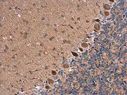
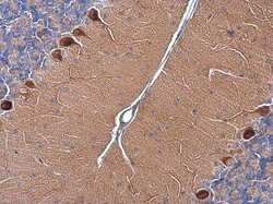
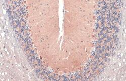
- Immunohistochemistry (Paraffin) analysis of RAB11B was performed in paraffin-embedded rat brain tissue using RAB11B Polyclonal Antibody (Product # PA5-31348) at a dilution of 1:500.
- Submitted by
- Invitrogen Antibodies (provider)
- Main image
- Experimental details
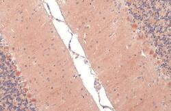
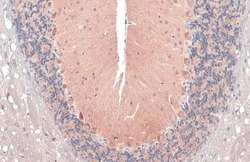
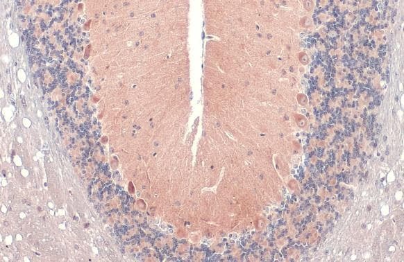
- Immunohistochemistry (Paraffin) analysis of RAB11B was performed in paraffin-embedded mouse brain tissue using RAB11B Polyclonal Antibody (Product # PA5-31348) at a dilution of 1:1000. Antigen Retrieval: Citrate buffer, pH 6.0, 15 min.

- Submitted by
- Invitrogen Antibodies (provider)
- Main image
- Experimental details
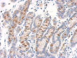
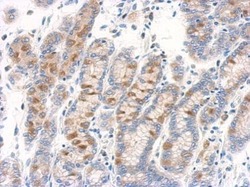
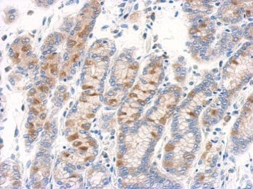
- Immunohistochemical analysis of paraffin-embedded human colon carcinoma, using RAB11B antibody (Product # PA5-31348) antibody at 1:500 dilution. Antigen Retrieval: EDTA based buffer, pH 8.0, 15 min.
- Submitted by
- Invitrogen Antibodies (provider)
- Main image
- Experimental details
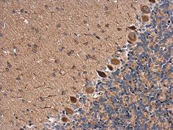
- RAB11B Polyclonal Antibody detects RAB11B protein at cytoplasm by immunohistochemical analysis. Sample: Paraffin-embedded mouse brain. RAB11B stained by RAB11B Polyclonal Antibody (Product # PA5-31348) diluted at 1:500. Antigen Retrieval: Citrate buffer, pH 6.0, 15 min.

- Submitted by
- Invitrogen Antibodies (provider)
- Main image

- Experimental details
- RAB11B Polyclonal Antibody detects RAB11B protein at cytoplasm by immunohistochemical analysis. Sample: Paraffin-embedded mouse brain. RAB11B stained by RAB11B Polyclonal Antibody (Product # PA5-31348) diluted at 1:500. Antigen Retrieval: Citrate buffer, pH 6.0, 15 min.

- Submitted by
- Invitrogen Antibodies (provider)
- Main image
- Experimental details
- RAB11B Polyclonal Antibody detects RAB11B protein at cytoplasm by immunohistochemical analysis. Sample: Paraffin-embedded rat brain. RAB11B stained by RAB11B Polyclonal Antibody (Product # PA5-31348) diluted at 1:500. Antigen Retrieval: Citrate buffer, pH 6.0, 15 min.

- Submitted by
- Invitrogen Antibodies (provider)
- Main image
- Experimental details
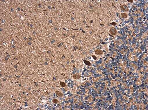
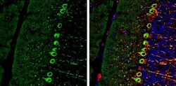
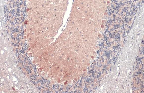
- Immunohistochemistry (Frozen) analysis of RAB11B was performed in frozen-sectioned adult mouse cerebellum tissue using RAB11B Polyclonal Antibody (Product # PA5-31348) at a dilution of 1:250 (Green). Red: NF-H, stained by NF-H antibody diluted at 1:500. Blue: Fluoroshield with DAPI. Antigen Retrieval: Citrate buffer, pH 6.0, 10 min.

- Submitted by
- Invitrogen Antibodies (provider)
- Main image
- Experimental details
- Immunohistochemistry (Paraffin) analysis of RAB11B was performed in paraffin-embedded rat brain tissue using RAB11B Polyclonal Antibody (Product # PA5-31348) at a dilution of 1:500.

- Submitted by
- Invitrogen Antibodies (provider)
- Main image

- Experimental details
- Immunohistochemistry (Paraffin) analysis of RAB11B was performed in paraffin-embedded mouse brain tissue using RAB11B Polyclonal Antibody (Product # PA5-31348) at a dilution of 1:1000. Antigen Retrieval: Citrate buffer, pH 6.0, 15 min.

- Submitted by
- Invitrogen Antibodies (provider)
- Main image
- Experimental details
- Immunohistochemical analysis of paraffin-embedded human colon carcinoma, using RAB11B antibody (Product # PA5-31348) antibody at 1:500 dilution. Antigen Retrieval: EDTA based buffer, pH 8.0, 15 min.
- Submitted by
- Invitrogen Antibodies (provider)
- Main image
- Experimental details
- RAB11B Polyclonal Antibody detects RAB11B protein at cytoplasm by immunohistochemical analysis. Sample: Paraffin-embedded mouse brain. RAB11B stained by RAB11B Polyclonal Antibody (Product # PA5-31348) diluted at 1:500. Antigen Retrieval: Citrate buffer, pH 6.0, 15 min.
- Submitted by
- Invitrogen Antibodies (provider)
- Main image

- Experimental details
- RAB11B Polyclonal Antibody detects RAB11B protein at cytoplasm by immunohistochemical analysis. Sample: Paraffin-embedded mouse brain. RAB11B stained by RAB11B Polyclonal Antibody (Product # PA5-31348) diluted at 1:500. Antigen Retrieval: Citrate buffer, pH 6.0, 15 min.
Supportive validation
- Submitted by
- Invitrogen Antibodies (provider)
- Main image
- Experimental details
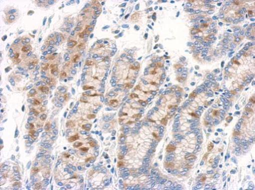
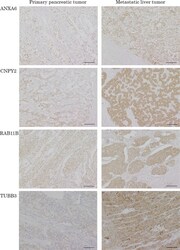
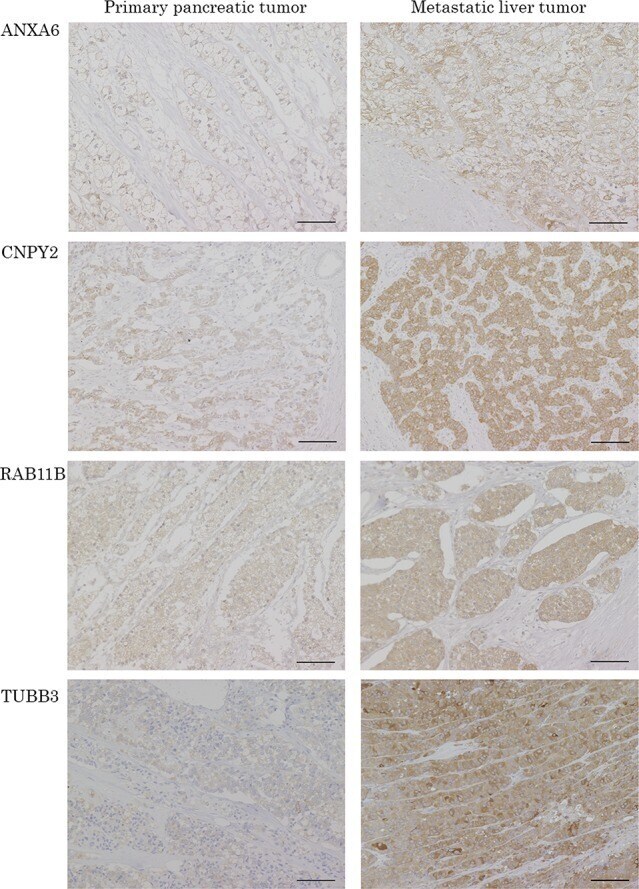
- Figure 3 Representative pictures of IHC for the candidate proteins in PT and paired LT The protein expressions of ANXA6, CNPY2, RAB11B and TUBB3 were confirmed in the patients evaluated with proteomics by IHC. ANXA6, CNPY2, RAB11B and TUBB3 are IHC images of patient NO.6, 4, 2 and 3, respectively. Scale bars indicate 100 mum.
- Submitted by
- Invitrogen Antibodies (provider)
- Main image
- Experimental details
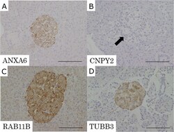
- Figure 4 Representative pictures of IHC for the candidate proteins in normal pancreatic tissue (A) ANXA6, (B) CNPY2, (C) RAB11B, (D) TUBB3. Expression of CNPY2 was negative in normal islet cells (B: arrow). ANXA6, RAB11B and TUBB3 had positive expression in normal islet cells. Expression of ANXA6, CNPY2, RAB11B and TUBB3 was not detected in normal exocrine tissue of the pancreas. Scale bars indicate 100 mum.

- Submitted by
- Invitrogen Antibodies (provider)
- Main image
- Experimental details
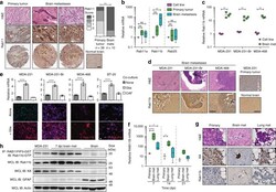
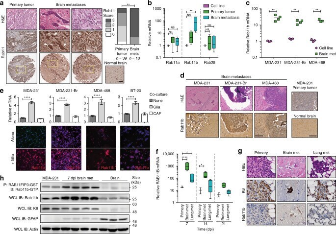
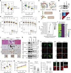
- Fig. 2 Rab11b is up-regulated during metastatic adaptation to the brain microenvironment. a Representative images of H&E (top) or Rab11 immunohistochemical staining of human primary breast cancer or breast cancer brain metastases (arrowheads). Left, Rab11 IHC scoring. Analysis of contingency, Fisher's exact test. Scale bar 100 mum. b qPCR for Rab11 isoforms in MDA-231 cells grown in culture, primary tumors (21 dpi), or brain metastases (21 dpi). Values for each isoform normalized to cells in culture. n = 3 independent cell samples, 7 animals/group. Boxes, first to third interquartile range, line, mean, whiskers, minimum and maximum values. ANOVA, Dunnett's multiple comparison. c qPCR for Rab11b in cells in culture versus brain metastases. Values are normalized to MDA-231 cells in culture. n = 3 independent cell samples, 4 animals/group Line, mean. Student's t test. d Representative H&E and Rab11b immunohistochemical staining of brain metastases, and MDA-231 primary tumor or murine brain. Scale bar 100 mum. e Top, qPCR for Rab11b in cell lines co-cultured with primary murine glia for 2 days. All values normalized to single culture. Bars, mean +- s.d. ANOVA, Dunnett's multiple comparison. Bottom, immunostaining for Rab11b (green, red) and nuclei (DAPI, blue) in cell lines cultured alone or co-cultured with primary murine glia for 5 days. n = 3. Scale bar 50 mum. f qPCR for Rab11b in MDA-231 primary tumors or metastases as indicated, collected at time points indicated. All value
- Submitted by
- Invitrogen Antibodies (provider)
- Main image
- Experimental details
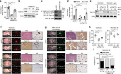
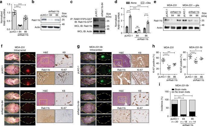
- Fig. 3 Rab11b is required for breast cancer brain metastasis. a Normalized mean RAB11B expression in MDA-231 cells, relative to pLKO.1 empty vector. Three independent experiments. Bars, mean +- s.d. ANOVA, Dunnett's multiple comparison. b Rab11b immunoblots for MDA-231 cells expressing indicated constructs. c Rab11b-GTP immunoblot for MDA-231 cells. d Normalized RAB11B expression in MDA-231 cells cultured alone or with primary murine glia for three days, relative to pLKO.1 alone. n = 3 independent experiments. Bars, mean +- s.d. Two-way ANOVA, Tukey's multiple comparison. e Rab11b immunoblots for MDA-231 cells cultured alone or with primary murine glia for five days followed by removal of glial cells. f Representative H&E and IHC images for mice intracranially injected with MDA-231-tdTomato control or shRab11b cells. Scale bar 100 mum. g Representative H&E and IHC images for mice intracardially injected with MDA-231-Br-EGFP control or shRab11b cells. Scale bar 100 mum. h Quantitation of Ki-67 staining. n = 2 independent experiments. Bars, mean +- s.d. ANOVA, Dunnett's multiple comparison. i Incidence of MDA-231-Br brain metastasis determined by visible GFP signal at 28 dpi. Analysis of contingency, Fisher's exact test. For all panels, * p < 0.05, ** p < 0.01, *** p < 0.001, **** p < 0.0001.
- Submitted by
- Invitrogen Antibodies (provider)
- Main image
- Experimental details
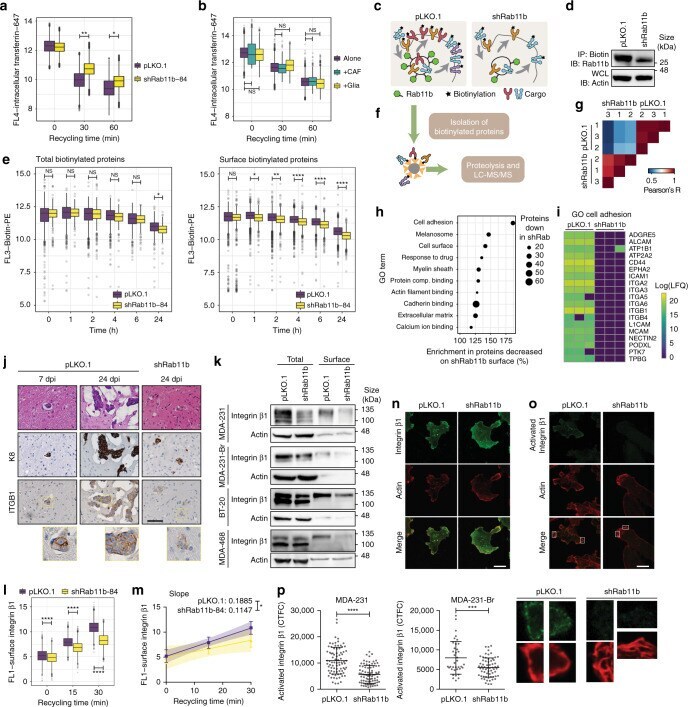
- Fig. 4 Rab11b recycling alters the surface proteome and controls integrin beta1 localization and activation. a Transferrin receptor recycling in MDA-231 cells. n = 3 independent experiments. Two-way ANOVA, Sidak's multiple comparison. b Transferrin receptor recycling in MDA-231 cells co-cultured for two days. Cancer cells were selected on expression of CD-44. n = 3 independent experiments. Two-way ANOVA, Sidak's multiple comparison. c Schematic of surface biotinylation. d Association of Rab11b with biotinylated surface proteins determined by immunoprecipitation and immunoblotting. e Retention of total or surface biotin following surface biotinylation with biotin-PE. n = 2 independent experiments. Two-way ANOVA, Sidak's multiple comparison. f Schematic of biotinylated surface protein isolation and proteomics. g Correlation matrix of all measured samples based on Pearson's correlation values. h Cleveland plot of top GO terms enriched in proteins that were decreased on the surface of shRab11b cells. i Heatmap of proteins annotated with GO term cell adhesion, containing at least one predicted transmembrane domain. j Representative images of H&E and IHC for mice intracranially injected with MDA-231-tdTomato cells. IHC for cytokeratin 8 (K8), Rab11b and integrin beta1 (ITGB1). Scale bar 50 mum. k Immunoblotting of total and surface lysates. l Surface integrin beta1 recycling in MDA-231 cells. n = 2 independent experiments. Two-way ANOVA, Sidak's multiple comparison. m Linear regres
- Submitted by
- Invitrogen Antibodies (provider)
- Main image
- Experimental details
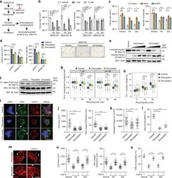
- Fig. 6 Statins decrease Rab11b localization and function. a Schematic of the mevalonate pathway leading to Rab11b geranylgeranylation. b Quantification of cells grown in soft agar. Bottom, representative images. Scale bar 1 mm. c Quantification of colony size and number for MDA-231 cells grown in soft agar with 1 muM pitavastatin/simvastatin, with 100 muM mevalonic acid or 10 muM geranylgeranylpyrophosphate. d Quantification of MDA-231 cells grown in soft agar with vehicle or 1 muM pitavastatin or simvastatin. e MDA-231 cells grown with vehicle or 10 muM-100 nM pitavastatin/simvastatin for 24 h. Immunoblotting of soluble and insoluble fractions separated with Triton X-114. f Rab11b activation assay for MDA-231 cells treated with vehicle or 1 muM pitavastatin/simvastatin for 24 h, followed by immunoblotting. g Transferrin receptor recycling in MDA-231 cells treated with vehicle or 1 muM pitavastatin or simvastatin. h Surface integrin beta1 recycling in MDA-231 cells treated with vehicle or 1 muM pitavastatin/simvastatin. ( i - l ) MDA-231 cells treated with 1 muM pitavastatin or simvastatin and adhered to decellularized murine brain matrix for 48 h. (I) Representative images of actin (phalloidin, green) protrusions (arrowheads), active integrin beta1 (12G10, red), and nuclei (DAPI, blue). Scale bar 10 mum. j Quantification of cell area and longest dimension. k Corrected total cellular active integrin beta1 fluorescence (CTCF). l Quantification of EdU. m - o MDA-231 cells treat
