 Explore
 Validate
 Learn
Learn Western blot
 Immunocytochemistry
Antibody data
- Antibody Data
- Antigen structure
- References [2]
- Comments [0]
- Validations
- Immunocytochemistry [3]
- Immunohistochemistry [1]
- Other assay [1]
Submit
Validation data
Reference
Comment
Report error
- Product number
- PA5-21541 - Provider product page

- Provider
- Invitrogen Antibodies
- Product name
- GALNT2 Polyclonal Antibody
- Antibody type
- Polyclonal
- Antigen
- Recombinant full-length protein
- Description
- Recommended positive controls: Molt-4, mouse adipose, rat brain. Predicted reactivity: Mouse (97%), Rat (98%), Zebrafish (92%), Rhesus Monkey (100%), Bovine (99%). Store product as a concentrated solution. Centrifuge briefly prior to opening the vial.
- Reactivity
- Human, Mouse, Rat
- Host
- Rabbit
- Isotype
- IgG
- Vial size
- 100 μL
- Concentration
- 1.77 mg/mL
- Storage
- Store at 4°C short term. For long term storage, store at -20°C, avoiding freeze/thaw cycles.
Submitted references The Golgi-associated retrograde protein (GARP) complex plays an essential role in the maintenance of the Golgi glycosylation machinery.
TLR7 in B cells promotes renal inflammation and Gd-IgA1 synthesis in IgA nephropathy.
Khakurel A, Kudlyk T, Bonifacino JS, Lupashin VV
Molecular biology of the cell 2021 Aug 15;32(17):1594-1610
Molecular biology of the cell 2021 Aug 15;32(17):1594-1610
TLR7 in B cells promotes renal inflammation and Gd-IgA1 synthesis in IgA nephropathy.
Zheng N, Xie K, Ye H, Dong Y, Wang B, Luo N, Fan J, Tan J, Chen W, Yu X
JCI insight 2020 Jul 23;5(14)
JCI insight 2020 Jul 23;5(14)
No comments: Submit comment
Supportive validation
- Submitted by
- Invitrogen Antibodies (provider)
- Main image
- Experimental details
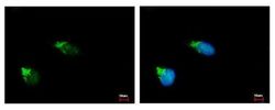
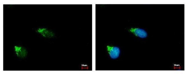
- Immunofluorescent analysis of GALNT2 showing staining in the Golgi apparatus of HeLa cells. HeLa cells were fixed in ice-cold MeOH for 5 min and stained using a GALNT2 polyclonal antibody (Product # PA5-21541) diluted at 1:500. Blue: Hoechst 33343 staining.
- Submitted by
- Invitrogen Antibodies (provider)
- Main image
- Experimental details
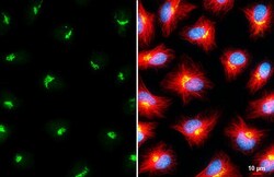
- GALNT2 Polyclonal Antibody detects GALNT2 protein at Golgi apparatus by immunofluorescent analysis. Sample: HeLa cells were fixed in ice-cold MeOH for 5 min. Green: GALNT2 stained by GALNT2 Polyclonal Antibody (Product # PA5-21541) diluted at 1:500. Red: alpha Tubulin, stained by alpha Tubulin Polyclonal Antibody [GT114] (Product # MA5-31466) diluted at 1:500. Blue: Fluoroshield with DAPI . Scale bar= 10 µm.

- Submitted by
- Invitrogen Antibodies (provider)
- Main image

- Experimental details
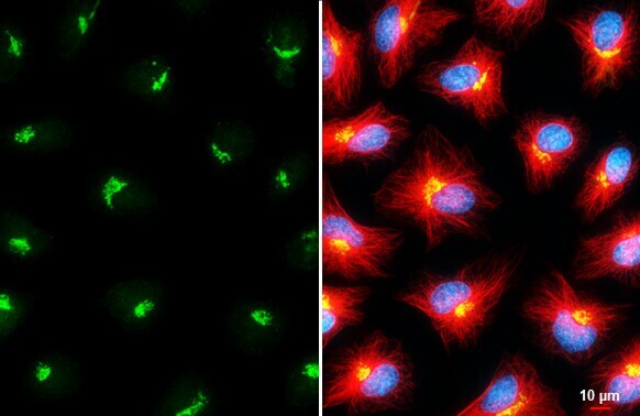
- GALNT2 Polyclonal Antibody detects GALNT2 protein at Golgi apparatus by immunofluorescent analysis. Sample: HeLa cells were fixed in ice-cold MeOH for 5 min. Green: GALNT2 stained by GALNT2 Polyclonal Antibody (Product # PA5-21541) diluted at 1:500. Red: alpha Tubulin, stained by alpha Tubulin Polyclonal Antibody [GT114] (Product # MA5-31466) diluted at 1:500. Blue: Fluoroshield with DAPI . Scale bar= 10 µm.
Supportive validation
- Submitted by
- Invitrogen Antibodies (provider)
- Main image
- Experimental details
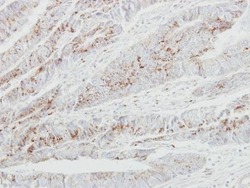
- Immunohistochemical analysis of paraffin-embedded human colon carcinoma, using GALNT2 (Product # PA5-21541) antibody at 1:250 dilution. Antigen Retrieval: EDTA based buffer, pH 8.0, 15 min.

Supportive validation
- Submitted by
- Invitrogen Antibodies (provider)
- Main image
- Experimental details
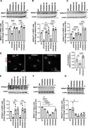
- FIGURE 3: GARP-KO affects the stability of key N- and O- Golgi glycosylation enzymes in RPE1 and HeLa cells. (A-C) WB (top panel) and quantification (bottom panels) of MGAT1 (A), B4GalT1 (B), and GalNacT2 (C) in WT, VPS53-KO, VPS54-KO, and the corresponding rescued RPE1 cells. (D) WT, VPS53-KO, and VPS53-KO rescued RPE1 cells were co-stained for endogenous B4GalT1 (green) and GM130 (magenta), and images were taken (left panel). Colocalization of B4GalT1 with GM130 was determined by calculation of the Pearson's correlation coefficient (right panel). At least 30 cells were imaged per sample for the quantification. Each dot in the bar graph (right panel) represents the colocalization of GM130 and B4GalT1 in several (1 to 10) cells imaged per field. (E) WB (top panel) and quantification (bottom panels) of ST6Gal1 in WT, VPS53-KO, VPS54-KO, and the corresponding rescued RPE1 cells. For quantification of ST6Gal1 blot, the additional low molecular weight band in VPS53-KO cells was not included. (F, G) WB (top panel) and quantification (bottom panels) of MGAT1 (F) and B4GalT1 (G) in WT, VPS50-, VPS51-, VPS52-, VPS53-, and VPS54-KO HeLa cells. Values in bar graphs represent the mean +- SD from three independent experiments. Statistical significance was calculated using one-way ANOVA. **** P
