Explore
 Validate
 Learn
Learn Western blot
 ELISA
ELISAAntibody data
- Antibody Data
- Antigen structure
- References [0]
- Comments [0]
- Validations
- Western blot [1]
- Immunocytochemistry [1]
- Immunohistochemistry [1]
Submit
Validation data
Reference
Comment
Report error
- Product number
- AP33452PU-N - Provider product page

- Provider
- OriGene
- Product name
- ARMETL1 (CDNF) rabbit polyclonal antibody, Aff - Purified
- Antibody type
- Polyclonal
- Description
- ARMETL1 (CDNF) rabbit polyclonal antibody, Aff - Purified
- Host
- Rabbit
- Conjugate
- Unconjugated
- Epitope
- CDNF
- Isotype
- IgG
- Antibody clone number
- NULL
- Vial size
- 100 µg
- Concentration
- 1.0 mg/ml
No comments: Submit comment
Supportive validation
- Submitted by
- OriGene (provider)
- Main image
- Experimental details
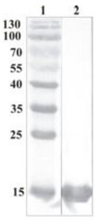
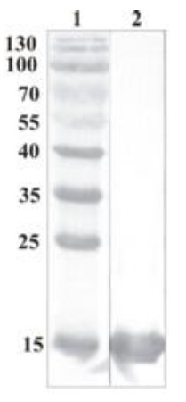
- Western Blot testing of anti-CDNF polyclonal antibody. Lane 1. PageRuler Prestained Protein Ladder ; Lane 2. Recombinant CDNF expressed into the supernatant of CHO cell culture medium.
- Validation comment
- WB
Supportive validation
- Submitted by
- OriGene (provider)
- Main image
- Experimental details
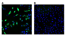
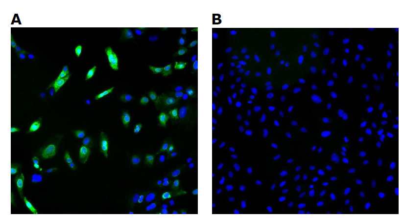
- Immunofluorescence detection of human CDNF expressed in U2OS cells. CDNF was visualized using anti-CDNF rabbit polyclonal antibody, dilution 1:3000. Goat ant-rabbit AlexaFluor488 was used as secondary antibody. For nuclear staining DAPI was used. ArrayScan VTI platform (Thermo Scientific) was used for image acquisition (10x objective). Composite picture was generated using pseudocolors green for CDNF specific signal and blue for nuclei. A. CDNF-expressing U2OS cells; B. Negative control (non-transfected U2OS cells).
- Validation comment
- IF
Supportive validation
- Submitted by
- OriGene (provider)
- Main image
- Experimental details
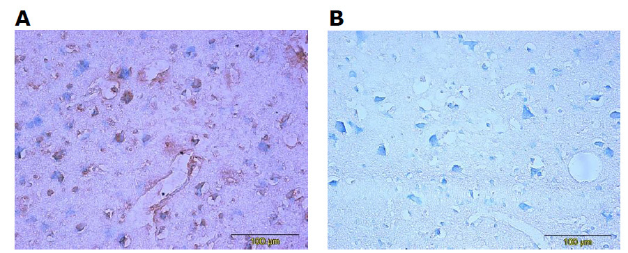
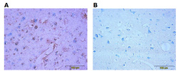
- Immunohistochemistry testing of anti-CDNF Rabbit polyclonal antibody. Analysis was performed using formalin-fixed paraffin-embedded human cerebral cortex tissue sections from Alzheimers disease patients. Tissue sections were boiled with sodium citrate buffer (pH 6) for antigen retrieval. Incubation with primary antibody at 5 ug/ml was performed overnight at 4°C. DAKO EnVisionTM Detection System, Peroxidase/DAB was used for visualization. Sections were counterstained with toluidine blue and mounted with Eukitt mounting medium. A. CDNF staining by rabbit polyclonal anti-CDNF antibody; B. Negative staining without primary antibody.
- Validation comment
- IHC