Explore
 Validate
 Learn
LearnPA5-27112
antibody from Invitrogen Antibodies
Targeting: DCLRE1C
A-SCID, ARTEMIS, FLJ11360, SCIDA, SNM1C
 Western blot
 Immunohistochemistry
Antibody data
- Antibody Data
- Antigen structure
- References [0]
- Comments [0]
- Validations
- Western blot [4]
- Chromatin Immunoprecipitation [1]
Submit
Validation data
Reference
Comment
Report error
- Product number
- PA5-27112 - Provider product page

- Provider
- Invitrogen Antibodies
- Product name
- Anti-Artemis Polyclonal Antibody
- Antibody type
- Polyclonal
- Antigen
- Recombinant protein fragment
- Description
- Recommended positive controls: A549, HeLa, HepG2, HCT116. Predicted reactivity: Rhesus Monkey (91%). Store product as a concentrated solution. Centrifuge briefly prior to opening the vial.
- Reactivity
- Human
- Host
- Rabbit
- Isotype
- IgG
- Vial size
- 100 µL
- Concentration
- 0.78 mg/mL
- Storage
- Store at 4°C short term. For long term storage, store at -20°C, avoiding freeze/thaw cycles.
No comments: Submit comment
Supportive validation
- Submitted by
- Invitrogen Antibodies (provider)
- Main image

- Experimental details
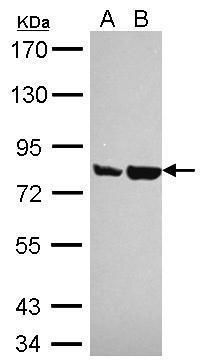
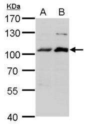
- Western blot analysis of Artemis using 30 µg of A) HeLa and B) HepG2 lysate. Samples were loaded onto a 7.5% SDS-PAGE gel and probed with an Artemis polyclonal antibody (Product # PA5-27112) at a dilution of 1:1000.
- Submitted by
- Invitrogen Antibodies (provider)
- Main image
- Experimental details
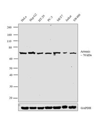
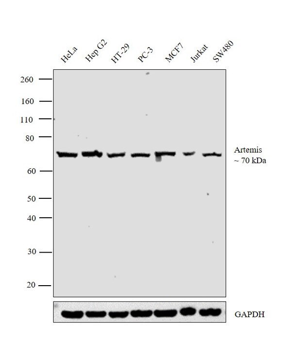
- Western blot analysis was performed on modified whole cell extracts (1% SDS) (30 µg lysate) of HeLa (Lane 1), Hep G2 (Lane 2), HT-29 (Lane 3), PC-3 (Lane 4), MCF7 (Lane 5), Jurkat (Lane 6) and SW480 (Lane 7). The blot was probed with Anti- Artemis Polyclonal Antibody (Product # PA5-27112, 1:1000 dilution) and detected by chemiluminescence using Goat anti Rabbit IgG (H+L) Superclonal™ Secondary Antibody, HRP conjugate (Product # A27036, 0.25 µg/mL, 1:4000 dilution). A 70 kDa band corresponding to Artemis was detected across the cell lines tested.
- Submitted by
- Invitrogen Antibodies (provider)
- Main image

- Experimental details
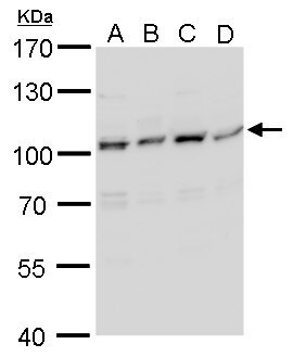
- Western Blot analysis of Artemis was performed by separating 30 µg of various whole cell lysates/extracts by 7.5 % SDS-PAGE. Proteins were transferred to a membrane and probed with a Artemis Polyclonal Antibody (Product # PA5-27112) at a dilution of 1:1000. A. A431, B. HeLa , C. HepG2 .
- Submitted by
- Invitrogen Antibodies (provider)
- Main image
- Experimental details
- Western Blot analysis of Artemis was performed by separating 30 µg of (A) MCF-7 and (B) MDA-MB-231 by 7.5 % SDS-PAGE. Proteins were transferred to a membrane and probed with a Artemis Polyclonal Antibody (Product # PA5-27112) at a dilution of 1:1000.

Supportive validation
- Submitted by
- Invitrogen Antibodies (provider)
- Main image
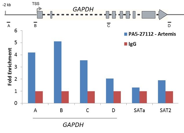
- Experimental details
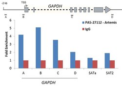
- Enrichment of endogenous Artemis protein at specific gene loci using Anti-Artemis Antibody: Chromatin Immunoprecipitation (ChIP) was performed using Anti-Artemis rabbit polyclonal antibody (Product # PA5-27112, 5 µg) on sheared chromatin from 2 million HeLa cells treated with Camptothecin (10 uM, 2 hr) using the MAGnify ChIP System (Product # 49-2024). Normal Rabbit IgG was used as a negative IP control. The purified DNA was analyzed by qPCR with PCR primer pairs over the GAPDH gene (positive), and SAT alpha and SAT2 satellite repeats (negative). Schematic diagram of the GAPDH gene is shown on top of the figure. Data is presented as fold enrichment of the antibody signal versus the negative control IgG using the comparative CT method.