Explore
 Validate
 Learn
Learn Western blot
 Immunohistochemistry
Antibody data
- Antibody Data
- Antigen structure
- References [4]
- Comments [0]
- Validations
- Western blot [1]
Submit
Validation data
Reference
Comment
Report error
- Product number
- HPA029616 - Provider product page

- Provider
- Atlas Antibodies
- Proper citation
- Atlas Antibodies Cat#HPA029616, RRID:AB_10602413
- Product name
- Anti-DTNBP1
- Antibody type
- Polyclonal
- Description
- Polyclonal Antibody against Human DTNBP1, Gene description: dystrobrevin binding protein 1, Alternative Gene Names: BLOC1S8, DBND, Dysbindin, HPS7, My031, Validated applications: WB, IHC, Uniprot ID: Q96EV8, Storage: Store at +4°C for short term storage. Long time storage is recommended at -20°C.
- Reactivity
- Human
- Host
- Rabbit
- Conjugate
- Unconjugated
- Isotype
- IgG
- Vial size
- 100 µl
- Concentration
- 0.1 mg/ml
- Storage
- Store at +4°C for short term storage. Long time storage is recommended at -20°C.
- Handling
- The antibody solution should be gently mixed before use.
Submitted references The Proteome of BLOC-1 Genetic Defects Identifies the Arp2/3 Actin Polymerization Complex to Function Downstream of the Schizophrenia Susceptibility Factor Dysbindin at the Synapse
TheN-Ethylmaleimide-Sensitive Factor and Dysbindin Interact To Modulate Synaptic Plasticity
Immunofluorescence and fluorescent-protein tagging show high correlation for protein localization in mammalian cells
Quantitative Proteomic and Genetic Analyses of the Schizophrenia Susceptibility Factor Dysbindin Identify Novel Roles of the Biogenesis of Lysosome-Related Organelles Complex 1
Gokhale A, Hartwig C, Freeman A, Das R, Zlatic S, Vistein R, Burch A, Carrot G, Lewis A, Nelms S, Dickman D, Puthenveedu M, Cox D, Faundez V
The Journal of Neuroscience 2016;36(49):12393-12411
The Journal of Neuroscience 2016;36(49):12393-12411
TheN-Ethylmaleimide-Sensitive Factor and Dysbindin Interact To Modulate Synaptic Plasticity
Gokhale A, Mullin A, Zlatic S, Easley C, Merritt M, Raj N, Larimore J, Gordon D, Peden A, Sanyal S, Faundez V
The Journal of Neuroscience 2015;35(19):7643-7653
The Journal of Neuroscience 2015;35(19):7643-7653
Immunofluorescence and fluorescent-protein tagging show high correlation for protein localization in mammalian cells
Stadler C, Rexhepaj E, Singan V, Murphy R, Pepperkok R, Uhlén M, Simpson J, Lundberg E
Nature Methods 2013;10(4):315-323
Nature Methods 2013;10(4):315-323
Quantitative Proteomic and Genetic Analyses of the Schizophrenia Susceptibility Factor Dysbindin Identify Novel Roles of the Biogenesis of Lysosome-Related Organelles Complex 1
Gokhale A, Larimore J, Werner E, So L, Moreno-De-Luca A, Lese-Martin C, Lupashin V, Smith Y, Faundez V
The Journal of Neuroscience 2012;32(11):3697-3711
The Journal of Neuroscience 2012;32(11):3697-3711
No comments: Submit comment
Enhanced validation
- Submitted by
- Atlas Antibodies (provider)
- Enhanced method
- Recombinant expression validation
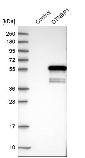
- Main image
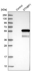
- Experimental details
- Western blot analysis in control (vector only transfected HEK293T lysate) and DTNBP1 over-expression lysate (Co-expressed with a C-terminal myc-DDK tag (~3.1 kDa) in mammalian HEK293T cells, LY403167).
- Sample type
- Human
- Protocol
- Protocol