Explore
 Validate
 Learn
Learn Western blot
 Immunohistochemistry
Antibody data
- Antibody Data
- Antigen structure
- References [1]
- Comments [0]
- Validations
- Immunohistochemistry [2]
- Other assay [4]
Submit
Validation data
Reference
Comment
Report error
- Product number
- PA5-20432 - Provider product page

- Provider
- Invitrogen Antibodies
- Product name
- ARMET Polyclonal Antibody
- Antibody type
- Polyclonal
- Antigen
- Synthetic peptide
- Description
- A suggested positive control is rat brain tissue lysate. PA5-20432 can be used with blocking peptide PEP-0549.
- Reactivity
- Human, Mouse, Rat
- Host
- Rabbit
- Isotype
- IgG
- Vial size
- 100 μg
- Concentration
- 1 mg/mL
- Storage
- Maintain refrigerated at 2-8°C for up to 3 months. For long term storage store at -20°C
Submitted references Bone marrow mesenchymal stem cells induce M2 microglia polarization through PDGF-AA/MANF signaling.
Yang F, Li WB, Qu YW, Gao JX, Tang YS, Wang DJ, Pan YJ
World journal of stem cells 2020 Jul 26;12(7):633-658
World journal of stem cells 2020 Jul 26;12(7):633-658
No comments: Submit comment
Supportive validation
- Submitted by
- Invitrogen Antibodies (provider)
- Main image
- Experimental details
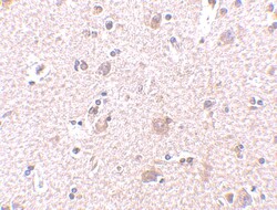
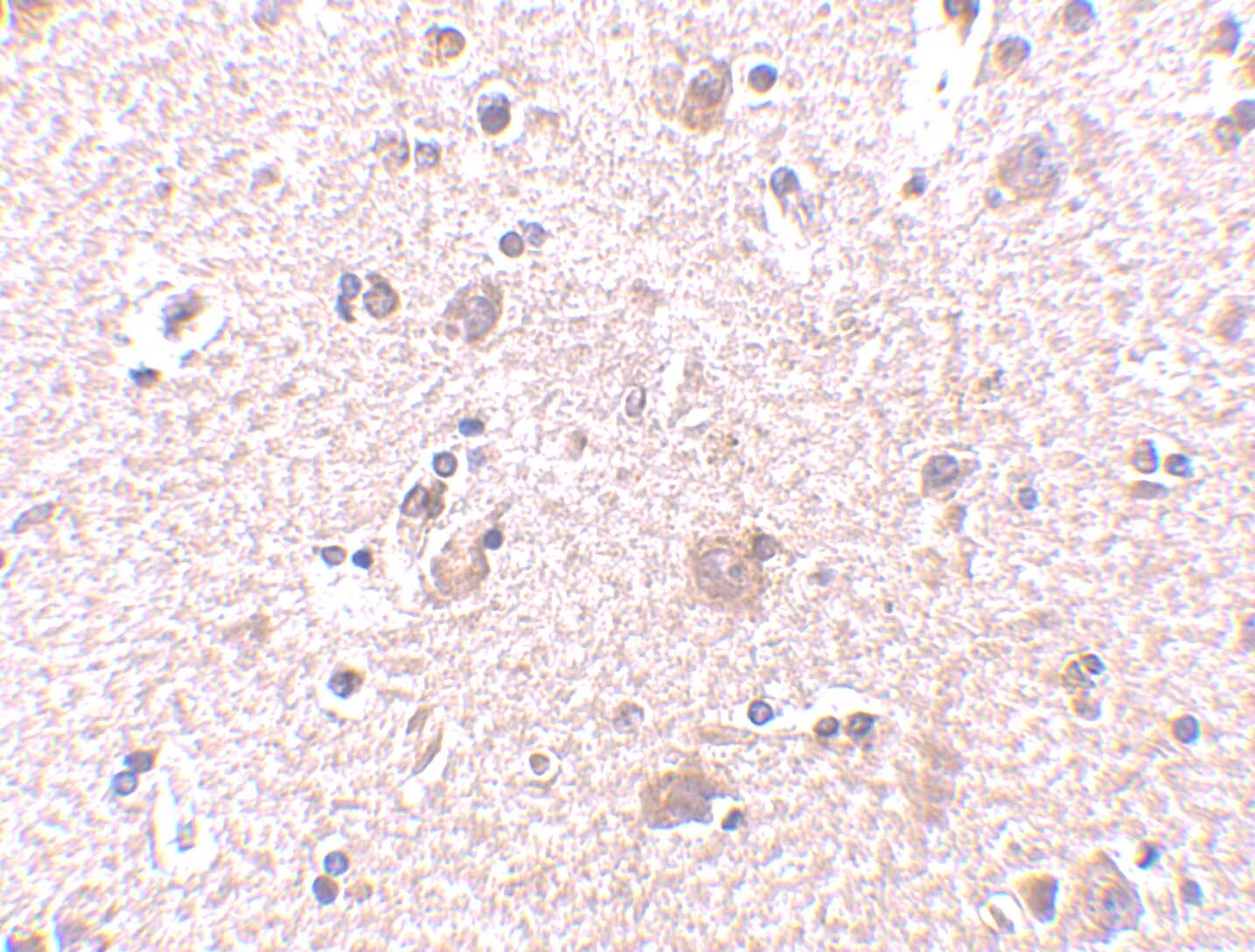
- Immunohistochemical analysis of paraffin-embedded human brain tissue using ARMET Polyclonal Antibody (Product # PA5-20432) at 2.5 µg /mL. Tissue was fixed with formaldehyde and blocked with 0.1 serum for 1 h at RT; antigen retrieval was by heat mediation with a citrate buffer (pH6). Samples were incubated with primary antibody overnight at 4˚C. A goat anti-rabbit IgG H&L (HRP) at 1/250 was used as secondary. Counter stained with Hematoxylin.
- Submitted by
- Invitrogen Antibodies (provider)
- Main image
- Experimental details
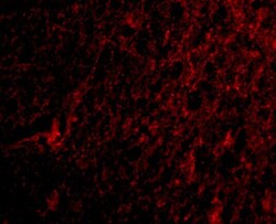
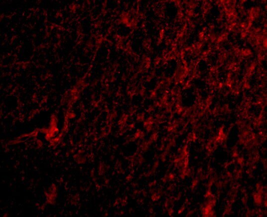
- Immunofluorescent analysis of 4% paraformaldehyde-fixed human brain tissue labeling MANF with ARMET Polyclonal Antibody (Product # PA5-20432) at 20 µg /mL, followed by goat anti-rabbit IgG secondary antibody at 1:500 dilution (red).
Supportive validation
- Submitted by
- Invitrogen Antibodies (provider)
- Main image
- Experimental details
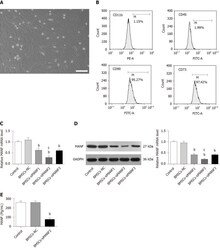
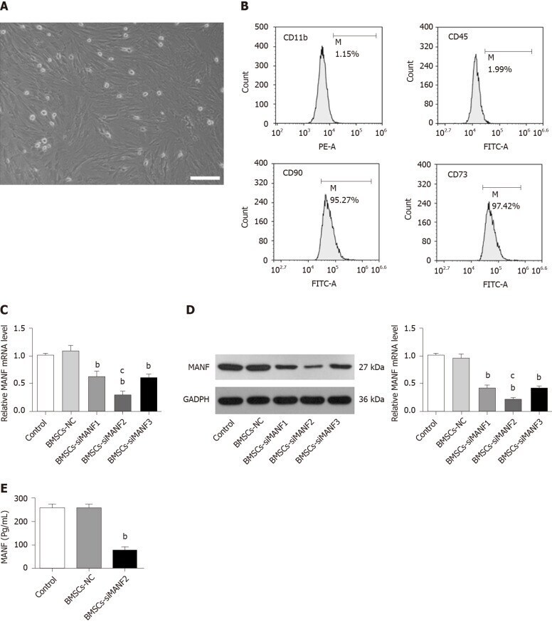
- Figure 2 C onstruction of genetically modified bone marrow mesenchymal stem cells. A: Cultured bone marrow mesenchymal stem cells (BMSCs) were homogeneous in size and morphology. Scale bar = 500 mum; B: Flow cytometry for detection of BMSCs surface markers; C and D: qRT-PCR (C) and Western blot analysis (D) of MANF levels in BMSCs transfected with MANF-siRNA or NC. E: ELISA of MANF in culture medium of BMSCs. b P < 0.01 vs Control and BMSCs-NC groups; c P < 0.05 vs BMSCs-siMNAF1 and BMSCs-siMANF3 groups. The values are expressed as the mean +- SD ( n =6). MANF: Mesencephalic astrocyte-derived neurotrophic factor; GAPDH: Glyceraldehyde-3-phosphate dehydrogenase; BMSCs: Bone marrow mesenchymal stem cells; BMSCs-NC: Negative control-transfected BMSCs; BMSCs-siMANF: MANF siRNA-transfected BMSCs.
- Submitted by
- Invitrogen Antibodies (provider)
- Main image
- Experimental details
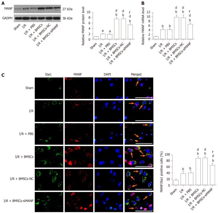
- Figure 4 Mesencephalic astrocyte-derived neurotrophic factor expression in microglia/macrophages in cerebral ischemia. A and B: Western blot (A) and qRT-PCR analysis (B) of MANF expression at 24 h after cerebral I/R injury; C: Immunohistochemical analysis of MANF expression in the microglia/macrophages of the injured brains. Representative images of immunohistochemical staining for the microglia/macrophage marker Iba1 (green) and MANF (red), with DAPI (blue) as a nuclear counterstain, are shown. a P < 0.05, b P < 0.01 vs Sham group; d P < 0.01 vs I/R and I/R + PBS groups; f P < 0.01 vs I/R + BMSCs and I/R + BMSCs-NC groups. Scale bar = 50 mum. Arrows point to the MANF + /Iba1 + cells. The values are expressed as the mean +- SD ( n = 6). MANF: Mesencephalic astrocyte-derived neurotrophic factor; GAPDH: Glyceraldehyde-3-phosphate dehydrogenase; I/R: Ischemia/reperfusion; PBS: Phosphate-buffered saline; BMSCs: Bone marrow mesenchymal stem cells; BMSCs-NC: Negative control-transfected BMSCs; BMSCs-siMANF: MANF siRNA-transfected BMSCs; Iba1: Ionized calcium-binding adapter molecule; DAPI: 4'6-diamidino-2-phenylindole.
- Submitted by
- Invitrogen Antibodies (provider)
- Main image
- Experimental details
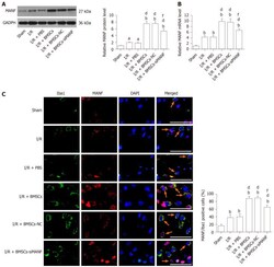
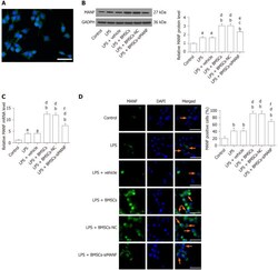
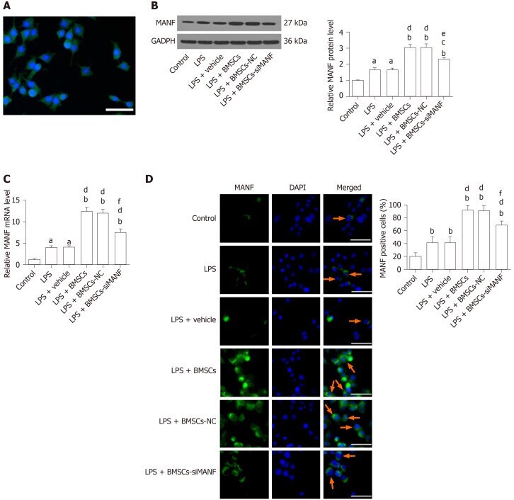
- Figure 5 Mesencephalic astrocyte-derived neurotrophic factor expression in lipopolysaccharide-stimulated microglia. A: Representative immunocytochemical staining for Iba1 (green) and co-staining for DAPI (blue) as a nuclear counterstain. Scale bar = 50 mum; B-D: Western blot (B), qRT-PCR (C), and immunocytochemistry analysis (D) of MANF expression in microglia pretreated with 100 ng/mL LPS for 24 h with or without BMSCs. Representative images of MANF (green), with DAPI (blue) as a nuclear counterstain, are shown. a P < 0.05, b P < 0.01 vs Control group; c P < 0.05, d P < 0.01 vs LPS and LPS + vehicle groups; e P < 0.05, f P < 0.01 vs LPS + BMSCs and LPS + BMSCs-NC groups. Scale bar = 50 mum. Arrows point to the MANF + cells. The values are expressed as the mean +- SD ( n = 6). MANF: Mesencephalic astrocyte-derived neurotrophic factor; GAPDH: Glyceraldehyde-3-phosphate dehydrogenase; LPS: Lipopolysaccharide; BMSCs: Bone marrow mesenchymal stem cells; BMSCs-NC: Negative control-transfected BMSCs; BMSCs-siMANF: MANF siRNA-transfected BMSCs; DAPI: 4'6-diamidino-2-phenylindole.
- Submitted by
- Invitrogen Antibodies (provider)
- Main image
- Experimental details
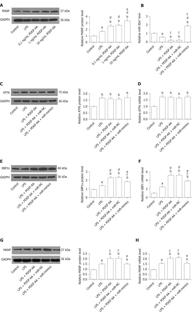
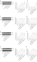
- Figure 9 Platelet-derived growth factor-AA-mediated induction of mesencephalic astrocyte-derived neurotrophic factor involves miR-30a*/X-box binding protein 1 signaling in HAPI cells stimulated by lipopolysaccharide. A: Western blot analysis of PDGF-AA-induced MANF expression in activated HAPI cells. a P < 0.05, b P < 0.01 vs Control group; d P < 0.01 vs LPS group; e P < 0.05 vs 0.1 ng/mL and 1 ng/mL PDGF-AA groups; B: qRT-PCR analysis of miR-30a* expression in untreated or 10 ng/mL PDGF-AA-treated activated HAPI cells transfected with or without miR-mimics or miR-NC; C and D: Western blot (C) and qRT-PCR analysis (D) of ATF6 expression in untreated or 10 ng/mL PDGF-AA-treated activated HAPI cells transfected with or without miR-mimics or miR-NC; E and F: Western blot (E) and qRT-PCR analysis (F) of XBP1 expression in untreated or 10 ng/mL PDGF-AA-treated activated HAPI cells transfected with or without miR-mimics or miR-NC; G and H: Western blot (G) and qRT-PCR analysis (H) of MANF expression in untreated or 10 ng/mL PDGF-AA-treated activated HAPI cells transfected with or without miR-mimics or miR-NC. a P < 0.05, b P < 0.01 vs Control group; c P < 0.05, d P < 0.01 vs LPS group; e P < 0.05, f P < 0.01 vs LPS + PDGF-AA and LPS + PDGF-AA + miR-NC groups. The values are expressed as the mean +- SD ( n = 6). MANF: Mesencephalic astrocyte-derived neurotrophic factor; PDGF-AA: Platelet-derived growth factor-AA; XBP1: X-box binding protein 1; ATF6: Activating transcription factor 6