Explore
 Validate
 Learn
Learn Western blot
Antibody data
- Antibody Data
- Antigen structure
- References [2]
- Comments [0]
- Validations
- Western blot [3]
Submit
Validation data
Reference
Comment
Report error
- Product number
- AF3748 - Provider product page

- Provider
- R&D Systems
- Product name
- Human MANF Antibody
- Antibody type
- Polyclonal
- Description
- Antigen Affinity-purified. Detects human MANF in direct ELISAs and Western blots.
- Reactivity
- Human
- Host
- Goat
- Conjugate
- Unconjugated
- Antigen sequence
P55145- Isotype
- IgG
- Vial size
- 100 ug
- Concentration
- LYOPH
- Storage
- Use a manual defrost freezer and avoid repeated freeze-thaw cycles. 12 months from date of receipt, -20 to -70 °C as supplied. 1 month, 2 to 8 °C under sterile conditions after reconstitution. 6 months, -20 to -70 °C under sterile conditions after reconstitution.
Submitted references Mesencephalic astrocyte-derived neurotrophic factor protects the heart from ischemic damage and is selectively secreted upon sarco/endoplasmic reticulum calcium depletion.
Mesencephalic astrocyte-derived neurotrophic factor is an ischemia-inducible secreted endoplasmic reticulum stress response protein in the heart.
Glembotski CC, Thuerauf DJ, Huang C, Vekich JA, Gottlieb RA, Doroudgar S
The Journal of biological chemistry 2012 Jul 27;287(31):25893-904
The Journal of biological chemistry 2012 Jul 27;287(31):25893-904
Mesencephalic astrocyte-derived neurotrophic factor is an ischemia-inducible secreted endoplasmic reticulum stress response protein in the heart.
Tadimalla A, Belmont PJ, Thuerauf DJ, Glassy MS, Martindale JJ, Gude N, Sussman MA, Glembotski CC
Circulation research 2008 Nov 21;103(11):1249-58
Circulation research 2008 Nov 21;103(11):1249-58
No comments: Submit comment
Supportive validation
- Submitted by
- R&D Systems (provider)
- Main image
- Experimental details
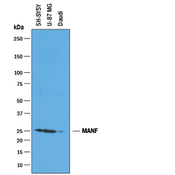
- Detection of Human MANF by Western Blot. Western blot shows lysates of SH-SY5Y human neuroblastoma cell line, U-87 MG human glioblastoma/astrocytoma cell line, and Daudi human Burkitt's lymphoma cell line. PVDF membrane was probed with 1 µg/mL of Goat Anti-Human MANF Antigen Affinity-purified Polyclonal Antibody (Catalog # AF3748) followed by HRP-conjugated Anti-Goat IgG Secondary Antibody (Catalog # HAF019). A specific band was detected for MANF at approximately 25 kDa (as indicated). This experiment was conducted under reducing conditions and using Immunoblot Buffer Group 1.

- Submitted by
- R&D Systems (provider)
- Main image
- Experimental details
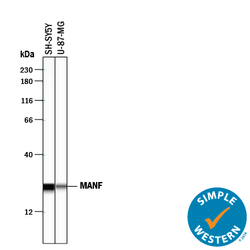
- Detection of Human MANF by Simple Western<SUP abp="261">TM. Simple Western lane view shows lysates of SH-SY5Y human neuroblastoma cell line and U-87 MG human glioblastoma/astrocytoma cell line, loaded at 0.2 mg/mL. A specific band was detected for MANF at approximately 24 kDa (as indicated) using 10 µg/mL of Goat Anti-Human MANF Antigen Affinity-purified Polyclonal Antibody (Catalog # AF3748) followed by 1:50 dilution of HRP-conjugated Anti-Goat IgG Secondary Antibody (Catalog # HAF109). This experiment was conducted under reducing conditions and using the 12-230 kDa separation system.

- Submitted by
- R&D Systems (provider)
- Main image

- Experimental details
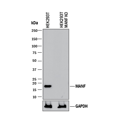
- Western Blot Shows Human MANF Specificity by Using Knockout Cell Line. Western blot shows lysates of HEK293T human embryonic kidney parental cell line and MANF knockout HEK293T cell line (KO). PVDF membrane was probed with 1 µg/mL of Goat Anti-Human MANF Antigen Affinity-purified Polyclonal Antibody (Catalog # AF3748) followed by HRP-conjugated Anti-Goat IgG Secondary Antibody (Catalog # HAF017). A specific band was detected for MANF at approximately 17 kDa (as indicated) in the parental HEK293T cell line, but is not detectable in knockout HEK293Tcell line. GAPDH (Catalog # AF5718) is shown as a loading control. This experiment was conducted under reducing conditions and using Immunoblot Buffer Group 1.