 Explore
 Validate
 Learn
Learn Western blot
 Immunocytochemistry
Antibody data
- Antibody Data
- Antigen structure
- References [2]
- Comments [0]
- Validations
- Immunocytochemistry [1]
Submit
Validation data
Reference
Comment
Report error
- Product number
- HPA015104 - Provider product page

- Provider
- Atlas Antibodies
- Proper citation
- Atlas Antibodies Cat#HPA015104, RRID:AB_1854173
- Product name
- Anti-MTDH
- Antibody type
- Polyclonal
- Description
- Polyclonal Antibody against Human MTDH, Gene description: metadherin, Alternative Gene Names: 3D3, AEG-1, LYRIC, Validated applications: WB, IHC, ICC, Uniprot ID: Q86UE4, Storage: Store at +4°C for short term storage. Long time storage is recommended at -20°C.
- Reactivity
- Human
- Host
- Rabbit
- Conjugate
- Unconjugated
- Isotype
- IgG
- Vial size
- 100 µl
- Concentration
- 0.1 mg/ml
- Storage
- Store at +4°C for short term storage. Long time storage is recommended at -20°C.
- Handling
- The antibody solution should be gently mixed before use.
Submitted references Metadherin, p50, and p65 expression in epithelial ovarian neoplasms: an immunohistochemical study.
Tumor suppressive microRNA-375 regulates oncogene AEG-1/MTDH in head and neck squamous cell carcinoma (HNSCC)
Giopanou I, Bravou V, Papanastasopoulos P, Lilis I, Aroukatos P, Papachristou D, Kounelis S, Papadaki H
BioMed research international 2014;2014:178410
BioMed research international 2014;2014:178410
Tumor suppressive microRNA-375 regulates oncogene AEG-1/MTDH in head and neck squamous cell carcinoma (HNSCC)
Nohata N, Hanazawa T, Kikkawa N, Mutallip M, Sakurai D, Fujimura L, Kawakami K, Chiyomaru T, Yoshino H, Enokida H, Nakagawa M, Okamoto Y, Seki N
Journal of Human Genetics 2011;56(8):595-601
Journal of Human Genetics 2011;56(8):595-601
No comments: Submit comment
Supportive validation
- Submitted by
- Atlas Antibodies (provider)
- Main image
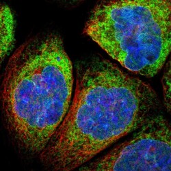
- Experimental details
- Immunofluorescent staining of human cell line A-431 shows localization to endoplasmic reticulum.
- Sample type
- Human
