 Explore
 Validate
 Learn
Learn Western blot
 ELISA
ELISA Immunoprecipitation
Antibody data
- Antibody Data
- Antigen structure
- References [0]
- Comments [0]
- Validations
- Western blot [1]
- Immunoprecipitation [2]
Submit
Validation data
Reference
Comment
Report error
- Product number
- LS-C396856 - Provider product page

- Provider
- LSBio
- Product name
- NDUFB6 Antibody LS-C396856
- Antibody type
- Polyclonal
- Description
- Protein G purified
- Reactivity
- Human
- Host
- Rabbit
- Isotype
- IgG
- Storage
- Short term: -20°C; Long term: -80°C; Avoid freeze-thaw cycles.
No comments: Submit comment
Enhanced validation
- Submitted by
- LSBio (provider)
- Enhanced method
- Genetic validation
- Main image
- Experimental details
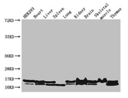
- Western Blot Positive WB detected in: HEK293 whole cell lysate, Mouse heart tissue, Mouse liver tissue, Mouse spleen tissue, Mouse lung tissue, Mouse kidney tissue, Mouse brain tissue, Mouse skeletal muscle tissue, Mouse thymus tissue All lanes: NDUFB6 antibody at 3µg/ml Secondary Goat polyclonal to rabbit IgG at 1/50000 dilution Predicted band size: 16, 14 kDa Observed band size: 16, 14 kDa

Supportive validation
- Submitted by
- LSBio (provider)
- Enhanced method
- Genetic validation
- Main image

- Experimental details
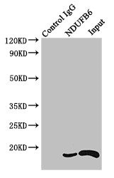
- Immunoprecipitating NDUFB6 in HEK293 whole cell lysate Lane 1: Rabbit monoclonal IgG (1µg) instead of NDUFB6 Antibody in HEK293 whole cell lysate.For western blotting, a HRP-conjugated Protein G antibody was used as the secondary antibody (1/2000) Lane 2: NDUFB6 Antibody (4µg) + HEK293 whole cell lysate (500µg) Lane 3: HEK293 whole cell lysate (20µg)
- Submitted by
- LSBio (provider)
- Main image

- Experimental details
- Immunoprecipitating NDUFB6 in HEK293 whole cell lysate Lane 1: Rabbit monoclonal IgG (1µg) instead of NDUFB6 Antibody in HEK293 whole cell lysate.For western blotting, a HRP-conjugated Protein G antibody was used as the secondary antibody (1/2000) Lane 2: NDUFB6 Antibody (4µg) + HEK293 whole cell lysate (500µg) Lane 3: HEK293 whole cell lysate (20µg)
